Introduction: the clinical challenge of upper airway obstruction (UAO)
UAO represents a significant hurdle in the success of non-invasive respiratory therapies (NIRT) and mechanical in-exsufflation (MIE).
In obstructive sleep apnea (OSA), available reports suggest that continuous positive airway pressure (CPAP) may accentuate the collapse of the epiglottis and render therapy ineffective and patients non-compliant.1,2
Patients with neuromuscular disorders (NMD) such as amyotrophic lateral sclerosis (ALS) may have OSA from the onset, with a prognostic impact.3 Moreover, when they are prescribed non-invasive ventilation (NIV), the frequency of UAO can reach up to 50%, primarily driven by bulbar dysfunction or REM-induced hypotonia.4,5
Another possible cause may be UAO induced by the oronasal mask, causing backward movement of the tongue.6
Importantly, studies indicate that ALS patients with non-corrected obstructive events on NIV have significantly reduced survival rates compared to those whose events are successfully managed.5
Pathophysiology and mechanisms of collapse
Understanding the specific site of collapse is crucial for adjusting therapy. The primary mechanisms include location-specific collapse (floppy epiglottis, soft palate collapse, oropharyngeal collapse, and paradoxical vocal fold movements) and therapy-induced (interface used, hyperventilation leading to glottic closure).
The interface used can have a significant influence on the occurrence of UAO. Over the years, studies have emerged demonstrating that the use of oronasal interfaces can be an inducing factor for UAO events, the resolution of which can often involve switching to a nasal interface, with the possible use of chin support to limit oral leakage.
One of the first descriptions of this phenomenon was made by Vrijsen et al.,6 in a patient with ALS, in whom it was found that the backward movement of the tongue during inspiration with an oronasal mask resulted in UAO. There has been a resolution of residual UAO through switching from an oronasal to a nasal interface in Duchenne muscular dystrophy (DMD).7 Studies in larger populations have also demonstrated the risk of obstructive events associated with the use of the oronasal interface,8,9 through mechanisms such as backward traction of the mandible induced by the mask and straps/harness, backward movement of the tongue (due to pressure applied to the mouth), reduction in the distance between the hyoid bone and the mandible, and shortening genioglossus muscle (since oronasal masks promote mouth breathing).9 It has even been suggested that switching from an oronasal to a nasal interface could be a first-line intervention, before considering further investigation such as polygraphy or transnasal fiberoptic videolaryngoscopy (TFL).9 However, nasal interfaces may have reduced effectiveness in patients with facial hypotonia.7
On the other hand, in one study, oronasal masks did not increase UAO,10 although ALS patients were not included, the subjects included required low EPAP levels, and patients and caregivers were strongly taught to avoid over-tightening of mask straps.
Beyond the interface issue, several pathophysiological mechanisms can give rise to UAO. ALS is a paradigmatic disease in this area, as in addition to causes common to other pathologies, it also includes causes linked to the involvement of the bulbar upper motor neuron.
In ALS, lability of the pharyngo-laryngeal muscles, characterized by spasticity and flaccidity, can lead to UAO.4 In Sarasate’s et al. study in ALS patients,4 it was shown that some of these individuals already present endoscopic changes in UA even during spontaneous ventilation (some of whom worsen with NIV), whereas others have been presenting changes during NIV, even without bulbar involvement at the time of evaluation (the authors suggest that bulbar involvement may already have existed before being detected by the scales).
There are several mechanisms that can cause UAO, particularly in ALS:
From a central point of view, we have bulbar dysfunction and instability of respiratory control, which leads to central apneas with glottic closure.4
From a mechanical point of view, we have weakness and hypotonia of the tongue and pharyngo-laryngeal musculature,4 which is a predisposing factor to increased upper airway collapsibility.11
Positive airway pressure can significantly increase cross-sectional dimensions at the level of the soft palate, lateral walls, and tongue base.12 On the other hand, NIV itself can also promote UAO events:
– Reduction of ventilatory drive and consequent passive glottic closure induced by reduced carbon dioxide levels.4,5 Sancho et al.13 studied ALS patients with UAO with decreased central drive (ODCD). They concluded that the vast majority of ODCD events were produced during non-rapid eye movement sleep stages, and that subjects with ODCDI > 5/h had upper motor neuron predominant dysfunction at the bulbar level and had greater controller gain and lower CO2 reserve.
– Reduction of ventilatory drive due to changes in thoracic afferents induced by NIV, independently of CO2 levels variation.4,5
– As a response of the upper airway to positive pressure, which includes adduction of the vocal cords (“glottic spasm”) and epiglottic drop (floppy epiglottis).4 In healthy subjects on NIV, increasing inspiratory positive airway pressure (IPAP) led to higher resistances in the upper respiratory tract, since the increase in resistance in the upper airway was greater than the increase in translaryngeal resistance, suggesting that high IPAP values affect the upper airway more extensively than the laryngeal structures.14 Furthermore, higher pressures increase the stress on flexible structures (particularly the most compliant ones, such as the pharynx and larynx), causing deformation and airway narrowing.14 High IPAP levels may aggravate epiglottic dysfunction, promoting occlusion of laryngeal spaces.15 In the case of ALS, upper motor neuron involvement causes hyperreflexia and hyperspasticity, increasing the risk of “glottic spasm,” as will be discussed later.
The obstruction can also be aggravated by retained oropharyngeal secretions and sialorrhea.11
In a mixed population (with predominance of obesity and COPD) of 13 subjects with persistent obstructions during NIV despite an EPAP of 12 cmH2O, Sayas Catalan et al.15 used TFL to identify the mechanism and the site of obstruction. They found out that obstruction under oronasal mask ventilation was due to soft-palate (velum) collapse in four subjects, to epiglottic backward movement in five subjects, and to tongue-base obstruction reducing the retroglossal space in three patients.
As discussed before, even in healthy people, resistance increases with elevated IPAP levels, particularly within the upper airway.14 In the study by Brekka et al.14 in healthy subjects under NIV, phenomena such as tendency for adduction of the aryepiglottic folds, high-rising epiglottis, retroflex epiglottis, and narrow hypopharynx were identified.
Another potential mechanism of UAO is related to expiratory obstruction at the level of the upper airway. Léotard et al.16 retrospectively analyzed 21 patients with DMD, and identified 4 cases of de expiratory obstructions, leading to end-inspiratory breath-holding and impossibility to take another inspiratory breath with a barometric mode until expiration occurs.
A summary of potential locations and mechanisms of UAO during NIV is presented in table 1.
Table 1. Potential locations and mechanisms of upper airway obstruction during NIV
| Type | Details |
|---|---|
| Central origin | Bulbar dysfunction and instability of respiratory control |
| Obstruction by collapse of the upper airway | Weakness and hypotonia of the tongue and pharyngo-laryngeal musculature |
| Induced by the interface (oronasal mask) | Backward traction of the mandible Backward movement of the tongue Reduction in the distance between the hyoid bone and the mandible shortens the genioglossus muscle |
| Induced by the ventilator settings/pressures | Reduction of ventilatory drive (due to reduction of PCO2) and consequent passive glottic closure Reduction of ventilatory drive due to changes in thoracic afferents Response of the upper airway to positive pressure Backward movement of the tongue Epiglottic drop (floppy epiglottis) Adduction of the vocal cords (“glottic spasm”) |
NIV: non-invasive ventilation.
MIE
With the use of TFL, it was observed that even in healthy individuals, various obstructive laryngeal movements can occur during the use of MIE, such as, for example, narrowing of the vocal folds, retroflexion of the epiglottis, hypopharyngeal constriction, backward movement of the base of the tongue,17 similar to what is observed with the use of NIV.
For several years, it has been demonstrated that patients with ALS and severe bulbar dysfunction exhibit a poorer response to MIE, with lower peak cough flow (PCF) values.18 Subsequently, studies using TFL showed adduction of supraglottic laryngeal structures during insufflation in bulbar ALS patients, as well as hypopharyngeal constriction during exsufflation in healthy and ALS (bulbar and non-bulbar) subjects, most prominently in patients with ALS and bulbar symptoms.19 Another study, also in ALS patients and using TFL, showed that laryngeal adduction occurred at lower insufflation pressures with disease progression. It was observed that retroflex movement of the epiglottis occurred in more than half of the patients, regardless of insufflation pressures and independent of bulbar involvement. A backward movement of the tongue was also observed in almost all patients (regardless of insufflation pressures) and constriction of the hypopharynx during exsufflation in all subjects, regardless of bulbar symptoms.20
From a pathophysiological point of view, bulbar upper motor neuron dysfunction is associated with obstruction during insufflation (upper motor neuron dysfunction results in spasticity and hyperreflexia, leading to laryngeal adduction), whereas bulbar lower motor neuron dysfunction is related to upper-airway collapse during exsufflation (lower motor neuron dysfunction results in oropharyngeal muscle weakness, hypotonia, and hyporeflexia, leading to collapse of supraglottic structures).21 Furthermore, the presence of alterations in the generated graphs depends on the severity of bulbar dysfunction21 (see the next section).
A summary of potential locations and mechanisms of UAO during MIE is presented in table 2.
Table 2. Potential locations and mechanisms of upper airway obstruction during MIE
| Insufflation | Exsufflation |
|---|---|
| Backward movement of the base of the tongue Retroflexion of the epiglottis Adduction of aryepiglottic folds Adduction or paradoxical movement of true vocal folds |
Hypopharyngeal constriction |
MIE: mechanical in-exsufflation.
Non-invasive monitoring and prediction tools
Before moving to invasive visualization, several tools can predict or identify UAO: ventilator data, flow-volume curves, and polysomnography (PSG) endotyping.
These tools are generally more easily accessible and less invasive for patients. First and foremost, we have the analysis of ventilator data. Modern software can detect residual UAO by identifying a decrease and flattening of inspiratory flow while inspiratory pressure remains stable.
However, the data provided by the ventilators may have limited accuracy. A recent state-of-the-art review from the SomnoNIV group,22 states that the reliability of the apnea-hypopnea index (AHI) provided by the ventilator’s built-in software varies depending on the manufacturer, with a tendency in some cases to underestimate the number of events. It was also highlighted that the AHI reliability is strongly dependent on leaks.
In centers with greater specialization, reading and interpreting flow-volume curves from the built-in software of NIV ventilators and MIE devices can identify UA closure qualitatively by showing an abrupt cessation of inspiratory and/or expiratory flow with sustained zero flow. However, this approach requires practice and experience on the part of the clinician, as well as the availability of time to analyze tracings from several nights.
The gold standard for non-invasive tools is polysomnographic endotyping, since polysomnographic airflow shapes can help predict the site of collapse.
UAO during NIV leads to a reduction in inspiratory flow, both in oropharyngeal and glottic etiologies.15
In a proposal from the SomnoNIV group23 UA obstructions during NIV were classified as UA with reduction of ventilatory drive, mixed events, or UA obstruction without reduction of ventilatory drive. The first type of events (reduction of ventilatory drive) was described as a progressive and smooth reduction in flow amplitude with unchanged amplitude of pressure signal, simultaneous reduction or disappearance of thoracic and abdominal belt signal, and switch of ventilator to back-up respiratory rate without thoracic or abdominal movements. UA obstructions without reduction of ventilatory drive were described as a sudden reduction in flow amplitude while maintaining inspiratory positive pressure (with pressure-controlled ventilators) and phase opposition or phase angle in thoracic and abdominal belts.
Op de Beeck et al.24 carried out a retrospective cohort study in patients with moderate-to-severe OSA, using PSG and DISE, having verified that a complete concentric collapse at the level of the palate and lateral wall collapse shared similar polysomnographic flow characteristics (skewed, scoopy), diametrically opposed to tongue base and epiglottis collapse.
Polysomnographic/polygraphic analysis is also essential for the correct characterization of expiratory obstructions of the upper airway.10
MIE
The data provided by MIE devices include adherence, inflation volume, and assisted PCF measured by the device’s internal pneumotachograph.25 High PCF values are desirable, ideally at least > 160 L/min,26 or even > 270 L/min.27 The lack of response in PCF at increasing pressures suggests that the patient presents some kind of intolerance to this pressure increase, namely, upper airway collapse.28
In MIE, flow waveform analysis plays a fundamental role, including verifying whether the PFC provided by the device corresponds to reality. Lacombe et al.28 showed that the transition between insufflation and exsufflation was associated with a 100 ms negative initial peak in flow waveform, corresponding to the compressible volume, so they considered PCF measurements reliable if the peak width was > 100 ms (if not, it could correspond to the compressible volume inside the limb). Active coughing maneuvers with poor synchronization between the patient and the MIE device increase the risk of confusing the true PCF with the compression volume.25 Furthermore, in the presence of upper airway collapse, the measured assisted PCF can be paradoxically high.29
Waveform analysis was effectively used to evaluate the effectiveness of MIE in patients with ALS,21 and it was found that a pattern of obstruction during insufflation occurred in 34 subjects (50.7%) and a pattern of upper-airway collapse during exsufflation occurred in 18 subjects (26%). Obstruction during insufflation was described as a spike at the beginning of insufflation in the flow-time graph (coinciding with a spike on the pressure-time curve during insufflation), with a subsequent flattening of the rest of the curve until the end of insufflation, in contrast with the smooth and concave stroke of the patterns described as normal.21 The pattern of upper-airway collapse during exsufflation corresponded to a flow decrease occurring abruptly after reaching PCF at the beginning of exsufflation.21
The usefulness of waveform analysis in MIE has also been evaluated in mixed populations with other NMD (including DMD and spinal cord injury),30 having ascertained that in patients with more pronounced respiratory muscle weakness, high-pressure MIE resulted in an increased rate of upper airway closure and patient discomfort. The vast majority of patients with evidence of upper airway collapse had DMD, about half had upper airway collapse only with high-pressure MIE, and most cases of occlusion occurred during exsufflation.
In the review by Chatwin et al.,25 a complete and updated methodology for analyzing MIE tracings is presented, giving particular emphasis to the flow curve (dependent variable). They suggest that the normal waveform should include an early positive peak and a decreasing exponential morphology after the peak during insufflation, while on exsufflation, there should be an early negative peak (corresponding to the PCF) and an increasing exponential morphology. In case of no leakage, the areas under the curve of inspired and expired flow should show minimal quantitative differences. The authors also mentioned that if the patient is instructed to cough during exsufflation, there will be one or more flow peaks of different magnitudes.
Regarding leakage, inspiratory leaks will result in a flattening or inversion of the inspiratory curve after the peak flow; also, in the case of inspiratory leak, the area under the inspiratory part will be clearly larger than the expiratory part.25
Regarding airway obstruction, the authors report that the various potentially implicated structures probably share a common semiology in waveforms, with a flattening of the flow waveform with a value close to zero, both inspiration and expiration. Waveform analysis also allows monitoring of the synchrony between the patient and the mechanically assisted cough device, enabling optimization of the insufflation and exsufflation time settings.25
The gold standard: TFL-figure
Direct visualization through fiberoptic endoscopy remains a primary diagnostic method to identify the mechanism of UAO during NIRT. Defining the exact setup and protocol is extremely important to standardize this procedure. Based on the published literature, we describe the setup for implementing TFL during NIRT (Fig. 1).4,14,15,17,19,20,31,32
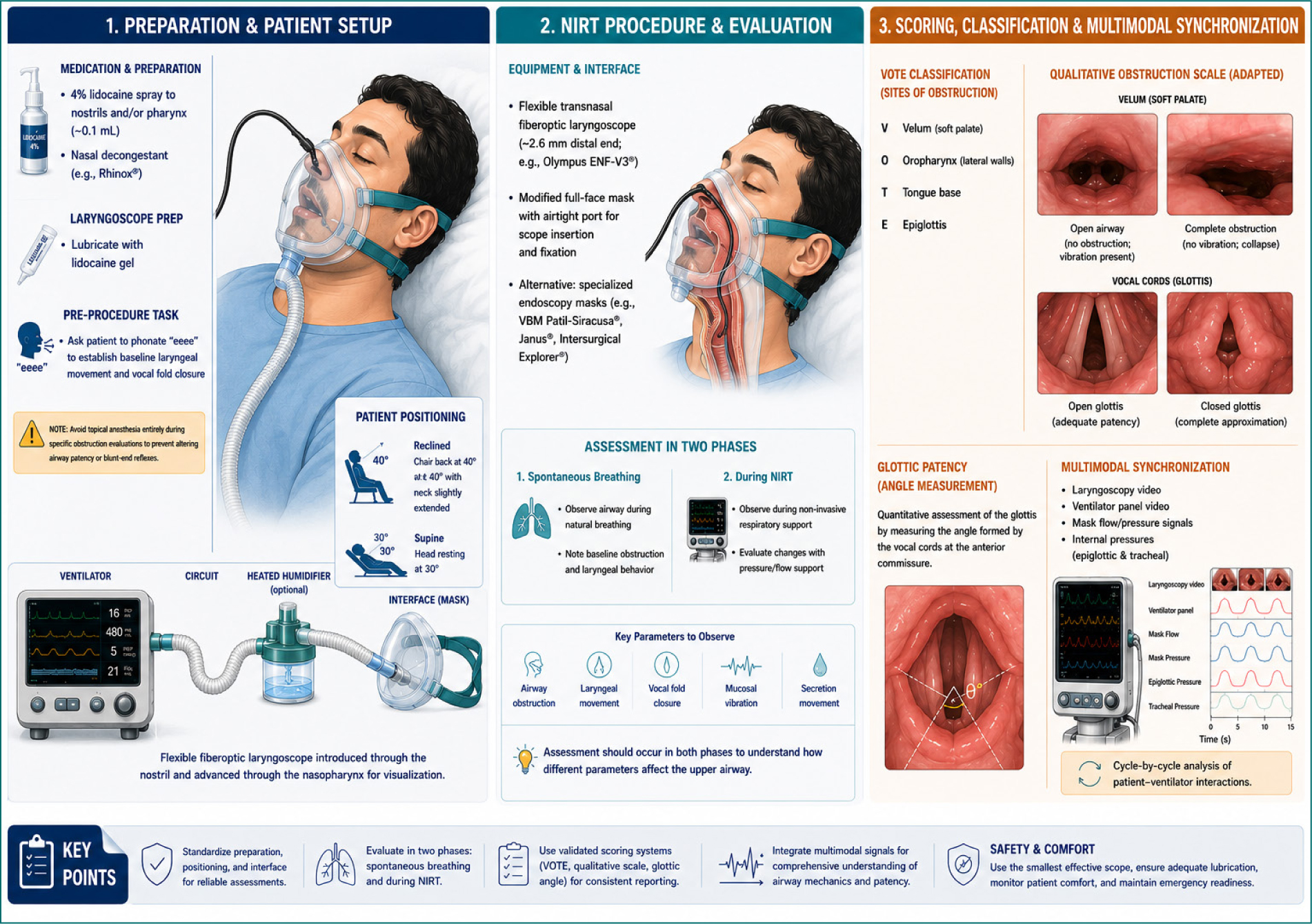
Figure 1. T setup of transnasal fiberoptic laryngoscopy during non-invasive respiratory therapies.
Medication and preparation
– Anesthesia: local anesthetic spray, typically 4% lidocaine, is applied to the nostrils and/or pharynx before insertion. In some protocols, a specific volume (e.g., 0.1 mL) is used.
– Lubrication: the laryngoscope should be lubricated with lidocaine gel to facilitate smooth entry through the nasopharynx.
– Decongestants: nasal decongestant sprays (e.g., Rhinox®) are often used to facilitate the introduction of the scope.
– Pre-procedure task: participants are typically asked to phonate “EEEE” to establish baseline laryngeal movement and vocal fold closure before therapeutic interventions begin.
Note: avoid topical anesthesia entirely during specific obstruction evaluations to prevent altering airway patency or blunt-end reflexes.
Patient positioning
– Reclined: a common standardized position is the chair back reclined at 40° with the neck slightly extended.
– Supine: some clinical protocols prefer the patient in a supine position with the head resting at 30°.
Laryngoscope
Flexible transnasal fiberoptic laryngoscopes with 2.6 mm distal end diameter are preferred for patient comfort and ease of use through masks (e.g., Olympus ENF-V3®).
Type of interface
– Modified full-face mask: a full facial mask is typically modified with a small airtight hole to allow for the insertion and fixation of the laryngoscope.
– The use of specialized endoscopy masks (e.g., VBM Patil-Siracusa®, Janus®, or Intersurgical Explorer®) may be an alternative.
Evaluation protocol
Assessment should occur in two phases: during spontaneous breathing and during NIRT, to observe how different parameters affect the airway.
Scoring and classification systems
There are several standardized methods for quantifying laryngeal behavior:
– VOTE classification: this evaluates four potential sites of obstruction:
1. Velum (soft palate)
2. Oropharynx (lateral walls)
3. Tongue base
4. Epiglottis
– Qualitative obstruction scale:
0: No obstruction (absence of vibration).
1: Partial obstruction (vibration).
2: Complete obstruction (collapse).
– Glottic patency (angle measurement): quantitative assessment of the glottis by measuring the angle formed by the vocal cords at the anterior commissure.
Multimodal synchronization
The most recent protocols14,31,32 integrate other simultaneous signals: laryngoscopy video, ventilator panel video, mask flow/pressure signals, and internal pressures (epiglottic and tracheal), allowing for cycle-by-cycle analysis of patient-ventilator interactions.
Emerging imaging and alternative techniques
Newer, less invasive technologies are proving useful for both clinical and research purposes: laryngeal ultrasound (US), magnetic resonance imaging (MRI), videofluoroscopy, and neck electrical impedance tomography (EIT).
Laryngeal US is emerging as a highly feasible, non-invasive alternative to TFL. It is significantly better tolerated by patients and shows a high concordance rate with TFL in observing laryngeal movements.
It is a technique that requires training and experience; however, it allows real-time visualization of upper airway structures, particularly at the glottis level. In the authors’ opinion, it is more easily performed during NIV than during MIE, since in the latter, the movements associated with coughing can lead to less efficient positioning of the probe during some phases of the cough cycle.
Larynx can be examined using US through an anterior view (to observe the vocal cords) by positioning the transducer transversely over the middle section of the thyroid cartilage, or by lateral incidence (allows visualization of the arytenoid cartilage movements, considered a surrogate measure of vocal folds movement), in which the transducer is placed laterally on both sides of the thyroid cartilage (vertically 1–1.5 cm inside and parallel to the lateral border of the thyroid cartilage, along the oblique line) to observe both arytenoid cartilages separately.33 However, the movements of vocal folds and aryepiglottic folds do not always respond similarly during NIV.33
Normal laryngeal movements during NIV should be abduction at the glottic and supraglottic levels during inspiration and adduction at both levels during expiration.33 However, even in healthy individuals, NIV-induced laryngeal obstruction, for example, inspiratory adduction of the vocal fold and/or aryepiglottic folds can occur.33
The reliability of laryngeal US (anterior and lateral approaches) was analyzed by Brekka et al.33 with TFL support: the visualization rate for all recorded breaths was 99.1% for TFL compared to 81.7% for laryngeal US; the discordance rate for the TFL versus laryngeal US observations was 11.1% for vocal fold movements and 11.7% for aryepiglottic fold movements. The visualization rate was slightly higher for the aryepiglottic folds than for the vocal folds. Three participants were only assessable with the lateral view (both lateral views). Most patients reported experiencing no discomfort during the US examination. The authors concluded that laryngeal US provided high-quality observations and high concordance with TFL and may serve as an alternative assessment when treatment failure is suspected to be of laryngeal origin.
Sancho et al.13 used US of the upper airway during NIV to determine the location of UAO with ODCD identified during PSG in ALS patients. They concluded that the vast majority of ODCD events were a consequence of an adduction of the vocal folds.
MIE
As for MIE waveform analysis, Chatwin et al.25 also proposed an algorithm for monitoring MIE using upper airway US, highlighting that, based on studies with TFL the upper airway responses to MIE may occur at the oropharynx (backward movement of the tongue base during insufflation and hypopharyngeal narrowing during exsufflation), supra-glottis (retroflex movement of epiglottis during insufflation and adduction of aryepiglottic folds during insufflation) and/or at the glottis (adduction or paradoxical movement of true vocal folds during insufflation). The oropharynx and hypopharyngeal areas can be visualized using a linear transducer placed in a transverse plane at the submandibular zone, midway between the mentum and hyoid bone, or using a curved low-frequency transducer placed submandibular (from the hyoid to the mentum) in a sagittal projection. At the supra-glottis level, it is possible to visualize the epiglottis using a linear transducer in a transverse plane at the thyrohyoid membrane. At the glottis level, a linear transducer placed at the thyroid cartilage in a transverse plane can be used to visualize the true vocal cords, vocal ligaments, and arytenoid cartilages (the latter can also be observed in a parasagittal view, parallel to the lateral border of thyroid cartilage).25
In the past, computed tomography scans of the upper airway were used to monitor MIE18 in patients with ALS, and it was observed a dynamic collapse of upper airway during exsufflation, with retraction of uvula and reduction of the lateral diameter of pharynx, mainly at the oropharynx (greater in patients with more severe bulbar dysfunction), however, this is a procedure that implies radiation exposure and the need to be performed in a proper room in radiology; also it is technically difficult to match the triggering of radiation and scanning with insufflation and exsufflation.25
MRI has been used previously to assess the effects of positive pressure on UA. Braga et al.34 found that the use of nasal CPAP in patients with sleep disorder breathing led to a consistent increase in functional residual capacity, and an increase in upper airway cross-sectional area at the level of the pharynx above the epiglottis at end expiration.
Some authors recommend that videofluoroscopy should be performed to assess UA during NIV in patients with low adherence to explore additional causal factors.35 Videofluoroscopy can also help to see the effect of different interfaces on the upper airway. Dorça et al.35 compared intranasal masks, oronasal masks, high-flow nasal cannula, and helmet CPAP in ALS patients and concluded that the greatest increase in pharyngeal cross-sectional area was observed with intranasal mask use, followed by high-flow nasal cannula, oronasal mask, and helmet, respectively.
Neck EIT may be a feasible tool for monitoring upper airway dynamics during sleep by measuring impedance variations in the sagittal plane. In fact, EIT was found to be a useful real-time monitoring device for detecting upper airway narrowing or collapse during natural sleep in patients with OSA, allowing for the estimation with good accuracy of changes in upper airway size.36
Piccin et al.37 compared, in OSA patients under CPAP with a nasal mask, the findings of EIT with those of TFL, and found that there was a high cross-correlation between flow and impedance curves, a high correlation between total neck impedance and velopharynx area, and a high correlation between total neck impedance and oropharynx area. Furthermore, inspiratory peak flow correlated with simultaneous neck impedance.
A summary of the advantages and disadvantages of the main techniques and tools used in visualization/monitoring of the upper airway is presented in table 3.
Table 3. Advantages and disadvantages of the main techniques and tools used in visualization/monitoring of the upper airway
| Technique/tool | Advantages | Disadvantages |
|---|---|---|
| Ventilator/MI device built-in software data | Easy access No extra costs Easy to interpret |
May have limited accuracy, particularly in the presence of leaks Risk of confusing the true PCF with the compressible volume |
| Flow waveform analysis | Easy access No extra costs Can identify UAO qualitatively Identify UAO during insufflation and/or exsufflation Identify leaks during MIE Optimize synchronization in MIE |
Requires time Requires expertise In MIE, common semiology in waveforms for different UAO mechanisms |
| Polygraphic/Polysomnographic endotyping | Non-invasive Differentiate between UAO with and without reduced ventilatory drive Polysomnographic airflow shapes can help predict the site of collapse |
Requires time Requires expertise Requires the availability of specific equipment and, in the case of polysomnography, an equipped sleep room |
| Transnasal fiberoptic videolaryngoscopy (TFL) | The gold standard for direct visualization of upper airway structures | Invasive procedure Requires time Requires expertise Requires the availability of specific equipment and, ideally, an endoscopy room |
| Upper airway ultrasound | Non-invasive No ionizing radiation Real-time visualization of upper airway structures High concordance rate with TFL Less uncomfortable than TFL Identify the location of UAO during MIE |
Requires time Requires expertise Requires the availability of specific equipment Operator dependent Increased difficulty during the use of MIE |
MIE: mechanical in-exsufflation; UAO: upper airway obstruction; PCF: peak cough flow.
Management and titration protocols
Once UAO is identified, it is necessary to make adjustments to the device parameters in order to try to counterbalance the pathophysiological mechanism.
Oropharyngeal/velum collapse typically requires an increase in EPAP to splint the airway. However, in the study by Sarasate et al.,4 there were ALS patients who did not improve from UAO with increased EPAP, and some even worsened with this increase.
In epiglottic collapse, the strategy may involve reducing both IPAP/EPAP and/or switching to a nasal interface to avoid high pressures applied through an oronasal interface. Sayas Catalan et al.15 improved persistent obstructions during NIV despite an EPAP of 12 cmH2O with oronasal mask by switching to nasal mask in 60% of subjects in a mixed population (using TFL for monitoring). In their protocol, data from built-in ventilator software were analyzed; in the presence of abnormal results, the patient underwent polygraphy/PSG with NIV titration; in case of residual events > 10/h despite EPAP > 12 cmH2O, TFL was performed.15
In situations of passive glottic closure (hyperventilation-induced), it is often necessary to decrease the IPAP (in order to reduce pressure support) to reduce rapid drops in PCO2 levels.
Other UAO correction protocols have been proposed. Georges et al.5 suggest increasing EPAP up to 10 cmH2O and subsequently switching to automatic mode, switching to volume-controlled mode, or trying other mechanical treatments. However, in a study by Panyarath et al.,38 automated modes (in comparison with fixed-pressure modes) were associated with higher AHI at 6 months, although the study was based on downloaded data from the ventilator. Sarasate et al.4 chose to increase EPAP (increments of 2 cmH2O up to 10 cmH2O), if obstruction persisted, PS was reduced in steps of 2 cmH2O but always kept at a minimum of 6 cmH2O. If obstruction continued, EPAP was then decreased, also in steps of 2 cmH2O, to a minimum of 4 cmH2O).
Imaging methods can also play an important role in the “live” titration of parameters.
The study by Sarasate et al.4 demonstrated the usefulness of UA endoscopy in resolving UAO events during NIV. In this study, the desired increase in lumen size (guided by TFL) was not achieved in only one patient. However, as already mentioned, endoscopy of the UAO is an invasive procedure that can be uncomfortable and carry risks.4
MIE
In the study by Sancho et al., an effective MIE was achieved in more than 98% of the patients after the parameters were adjusted based on flow waveform analysis.21 The authors propose the following protocol for ALS patients: in those with Norris bulbar subscore > 27, adjust MIE parameters according to tolerance and assisted PCF > 2,7 L/s; in those in Norris bulbar subscore < 27, adjust MIE parameters based on waveform analysis (change insufflation pressure and flow in case of obstruction during insufflation, change exsufflation pressure if obstruction during exsufflation, then adjust insufflation and exsufflation time).21 The obstructive events detected both in insufflation and exsufflation were corrected by decreasing the pressure setting (mean optimal parameters were insufflation pressure 34.25 ± 7.55 cmH2O, exsufflation pressure 39.62 ± 4.87 cmH2O, insufflation time 1.65 ± 0.44 s, exsufflation time 2.94 ± 0.20 s, and high insufflation flow). However, other authors suggest longer insufflation times, shorter exsufflation times, and slower insufflation flows.39,40
Chatwin et al.25 proposed an algorithm to titrate MIE setting using flow waveform analysis: initially, cycles with significant leaks should be discarded; then, if the waveform suggests inspiratory or expiratory closure, pressures should be reduced; subsequently, the insufflation and exsufflation times should be adjusted based on the flow levels at the transitions from insufflation to exsufflation and vice versa.
Pressure and flow titration under TFL monitoring is also important in MIE, as a reduction in insufflation pressures can help prevent adduction of supraglottic structures and promote laryngeal abduction.41 A recent study in healthy individuals31 with TFL monitoring supports using lower insufflation pressures and higher negative pressures in clinical practice, since positive pressures reached the trachea effectively, whereas negative tracheal pressures during exsufflation were approximately one-half of the intended settings.
Future directions: artificial intelligence (AI)
The field is moving toward AI-assisted awake endoscopic video analysis and AI-powered US for automated detection of OSA and site-specific collapse.42,43
In addition, surface electromyography recording of upper airway dilator muscles or flexible catheter-based sensor arrays could be used to monitor muscle activity or pressure directly on upper airway soft tissues.14,44
Conclusion and take-home messages
Visual assessment of the upper airway is indicated for all ALS patients and those with refractory events under NIV.
Minimum standards of TFL setup under NIV are highly recommended. However, while fiberoptic endoscopy is highly recommended, laryngeal US is a promising, better-tolerated alternative.
Ultimately, re-titrating NIV protocols based on the specific site of collapse is essential for improving patient outcomes and survival.
Funding
None.
Conflicts of interest
None.
Ethical considerations
Protection of human subjects and animals. The authors declare that no experiments on humans or animals were performed for this research.
Confidentiality, informed consent, and ethical approval. This study does not involve personal patient data, medical records, or biological samples, and does not require ethical approval. SAGER guidelines do not apply.
Declaration on the use of artificial intelligence. The authors declare that no generative artificial intelligence was used in the writing or creation of the content of this manuscript.
