Introduction
Chronic hypercapnic respiratory failure (CHRF) is characterized by sustained periods of decreased alveolar ventilation resulting in chronic diurnal increasing of the arterial partial pressure of carbon dioxide (PaCO2 > 45 mmHg). There are many mechanisms, isolated or in combination, involved in CHRF depending on the main causative disease: inadequacy of the respiratory drive, impaired neuronal transmission of the breathing impulses, pathological myopathy leading to inadequate respiratory muscular strength, abnormalities of the musculoskeletal system of the thorax, increased dead-space or severe airflow obstruction. Non-invasive ventilatory (NIV) support has shown to improve multiple outcomes such as quality of life, gas exchange, sleep disturbances, daytime symptoms, and to reduce hospitalizations and mortality.1–3 Adaptation to NIV is usually conducted during daytime that may not reflect changing sleep ventilatory patterns relying on sleep stages or nocturnal upper airway (UA) respiratory events or unintentional leaks, leading to unsuspected patient-ventilation asynchronies. Optimization of NIV is crucial and has been related to relevant outcomes, including survival.4,5 Nevertheless, the optimal titration method for long-term ventilated patients is still debated. Hence, the methods used to assess NIV efficacy may vary greatly, ranging from awake blood gas measurements to a sequence of full polysomnographic studies.
The American Academy of Sleep Medicine (AASM) recommended polysomnography (PSG) as the standard method for NIV titration in patients with hypoventilation and advised PSG confirmation when NIV was initiated empirically.6 Parameter adjustment via PSG has demonstrated significant advantages: (1) allows the detection of UA obstruction (UAO) events that may occur in case of the worsening of the patient’s condition, in the presence of unintentional leaks and/or caused by the interface; (2) incorporates respiratory effort based on thoraco-abdominal belts, enabling easily identification of the type of UAO events: with or without increased ventilatory drive; (3) awakening periods can be discarded and respiratory events can be detected according to the patient’s position and sleep stages and (4) making possible an accuracy interpretation of patient-ventilator asynchronies (PVA). However, there are several reasons why routinely PSG both for initiation and eventually re-titration/monitoring during follow-up is not feasible in our daily practice, ranging from the growing number of patients, which means that sleep units, already overwhelmed by long waiting lists, cannot cope, to the elevated costs associated, to the level of expertise and management skills required to carry out these studies. Thus, currently, the standard of care for NIV initiation relies on a pragmatical step-by-step approach.7,8 Recent statements propose algorithms for NIV titration during consecutive nights using a combination of daytime arterial blood gas analysis, nocturnal oximetry, capnography, and data derived from built-in monitoring systems of home ventilators. Several studies have demonstrated the reliability of these systems and suggested their potential use in clinical practice.9,10 Furthermore, actually, some software systems allow a breath-by-breath evaluation of flow and pressure, proposing raw signals close to those provided by sleep studies. This stepwise approach has also been validated for home NIV initiation.11,12 The systematic use of a basic screening combined with data from ventilator software could allow NIV to be optimised, limiting the indication of PSG to complex cases in which abnormalities persist after basic clinical and ventilatory assessments. For these reasons, this review aims to describe PSG-based ventilatory titration procedures and to provide evidence-based specific recommendations for the titration and monitoring of NIV using PSG.
PSG titration at initiation of NIV: signals, recommended procedure, and general evidence
Signals and procedure
The AASM describes requirements of the PSG-guided NIV titration6 as a comprehensive set of signals organized into two main categories: standard PSG signals channels (sleep staging/arousal) and respiratory/ventilatory channels (Fig. 1). Airflow signal could be obtained from: (1) via an external pneumotachograph inserted between the mask and a single-limb circuit, or the ventilator’s internal flow sensor integrated into the polysomnogram (used also to assess tidal volume, respiratory rate and leaks), or (2) through mask pressure signal (recorded via a pressure transducer at the mask to display the inspiratory positive airway pressure [IPAP]/expiratory positive airway pressure [EPAP] waveform). Alternative methods for assessing respiratory effort include diaphragmatic electromyography, diaphragmatic electrical activity, or esophageal manometry, which may also not be easily feasible in routine clinical practice.
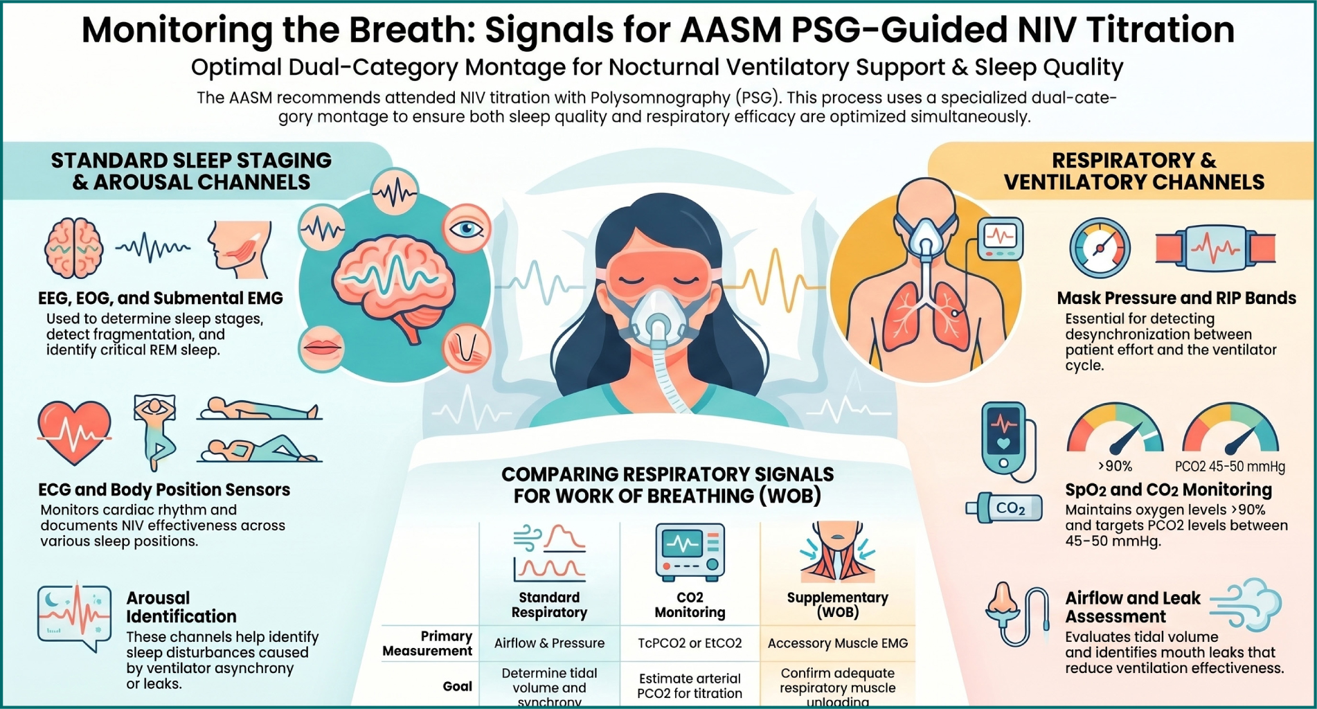
Figure 1. Signals during polysomnography under non-invasive non-invasive ventilation.
The optimal setting recommended for the nocturnal titration of NIV is in an accredited sleep center or laboratory with personnel experienced with both NIV titration and managing patients with hypoventilation, such as a registered PSG technologist. The titration should be reviewed and treatment pressures selected by a physician board certified in sleep medicine. Figure 2 summarizes the titration algorithm in accordance with the AASM’s recommendations.6,13 Recently, Berlowitz et al.14 had published a proposal for an attended overnight titration PSG in amyotrophic lateral sclerosis (ALS) patients. Their algorithm, pending on the completion of the trial to test the efficacy of its procedure, closely follows the recommendations set out by the AASM, with a few specific details. They propose to begin by initiating EPAP and IPAP 2 cm H2O lower than those established during the daytime acclimatation, increasing EPAP in 1 cm H2O whenever obstructive events were detected or if ineffective respiratory efforts occur without significant leak. The initial IPAP-EPAP gradient should be preserved until obstructive events are adequately controlled. When hypoventilation was identified, the primary intervention is to increase IPAP, raising pressure support. A 10 mmHg rise in transcutaneous CO2 above the awake, supine baseline should trigger such an adjustment. If the transcutaneous CO2 reading appears unreliable, the sensor should be reapplied and reassessed before any action is taken. Further increases in IPAP may also be tested to reduce non-obstructive hypopneas, alleviate flow limitation, or enhance oxygen saturation. If signs of partial UAO persist despite these measures, an additional incremental increase in EPAP should be trialled, while ensuring that pressure support remains constant (Fig. 3).
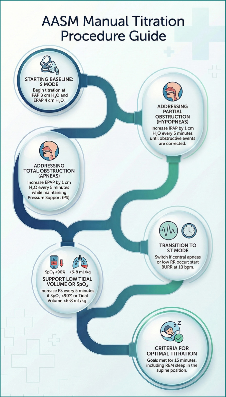
Figure 2. American Academy of Sleep Medicine guided-polysomnography titration of non-invasive ventilator.
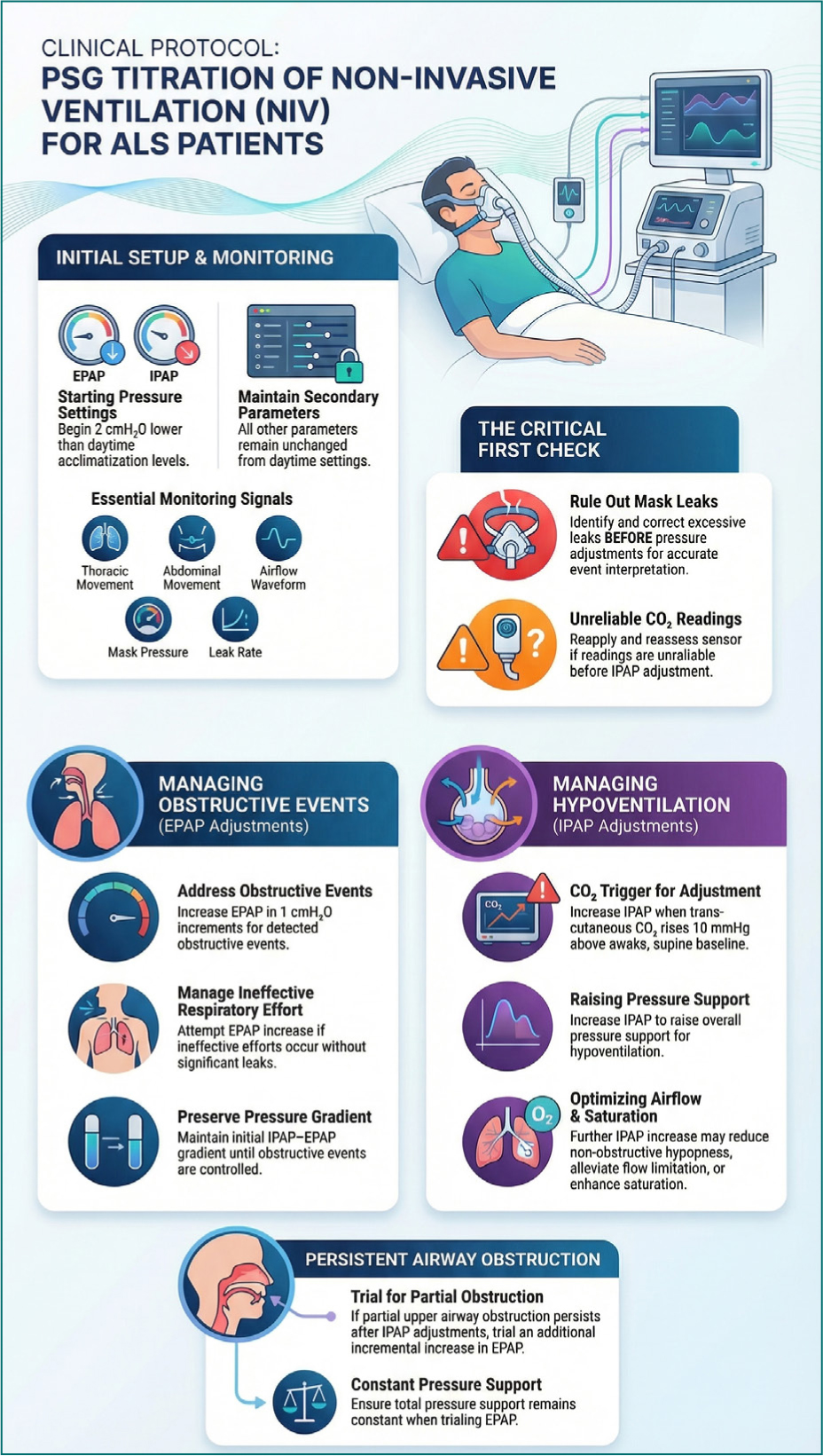
Figure 3. Proposal of guided-polysomnography titration of non-invasive ventilator in amyotrophic lateral sclerosis patients.
Pitfalls
An important point to consider is the impact of signal-filtering settings on the airflow signal during its acquisition on an attended PSG performed under NIV. In a recent scientific letter,15 the authors explain that, although signal filtering is essential for improving readability and reducing noise, the recommendations issued by the AASM for diagnostic sleep studies have not been validated for use in PSG under mechanical ventilation. Because the physiological signals captured during NIV differ substantially from those recorded in standard sleep studies, particularly due to the presence of intentional and unintentional leaks, the authors set out to determine whether these recommended filters may distort clinically relevant information. Using a library of clinical and bench recordings that included normal tracings, different types of leaks, airway obstruction, asynchronies, and artifacts, the authors evaluated the influence of various high-pass filter (HPF) and low-pass filter (LPF) cut-off points. Their analysis showed that HPF, even when applied with very low cut-off frequencies, frequently removed essential low-frequency components of the flow signal. As a result, important information regarding leaks was lost and, in some cases, misleading features resembling air trapping were artificially introduced, being most prominent in situations involving continuous, intermittent, or asymmetric leakage. By contrast, high-pass filtering had little effect on signals representing obstruction or patient-ventilator asynchrony. LPF, on the other hand, were generally safer: they did not significantly alter signals related to leaks, obstruction, or asynchronies. Their most relevant effect was in reducing high-frequency artifacts, although only filters with lower cutoffs (around 25 Hz) proved effective, whereas higher cutoffs recommended for snoring detection (50–100 Hz) did not adequately eliminate noise. The authors conclude that, for PSG performed under NIV, the routine use of HPFs should be avoided due to the risk of signal distortion and loss of key diagnostic information, particularly regarding leakage. They recommend the use of LPF with a cutoff near 25 Hz, which improves signal clarity without compromising waveform morphology. They further caution that some PSG devices incorporate non-removable HPF at the hardware level and may therefore be unsuitable for monitoring patients on NIV.
During polysomnographic assessment of NIV, temporal delays between ventilator signals and thoraco-abdominal effort recordings represent an important technical limitation that can affect the interpretation of patient-ventilator interaction (Fig. 4). These delays originate from several complementary mechanisms along the signal transmission and acquisition pathway. First, there is an inherent pneumatic delay, as pressure changes generated by the ventilator require a finite time to travel through the circuit and interface before producing a measurable mechanical response of the chest and abdomen. Second, differences in signal processing and sampling between the ventilator and the PSG system, such as varying sampling rates, filtering, and analog-to-digital conversion, can introduce additional offsets between pressure, flow, and effort signals.16 Finally, sensor-related factors contribute, as thoraco-abdominal belts have intrinsic response times that are slower than direct pressure measurements at the mask.17 Taken together, these factors can create small but clinically relevant temporal mismatches that must be considered when scoring and interpreting patient-ventilator asynchrony during PSG, to avoid confusing expected physiological lag with true trigger or cycling abnormalities.
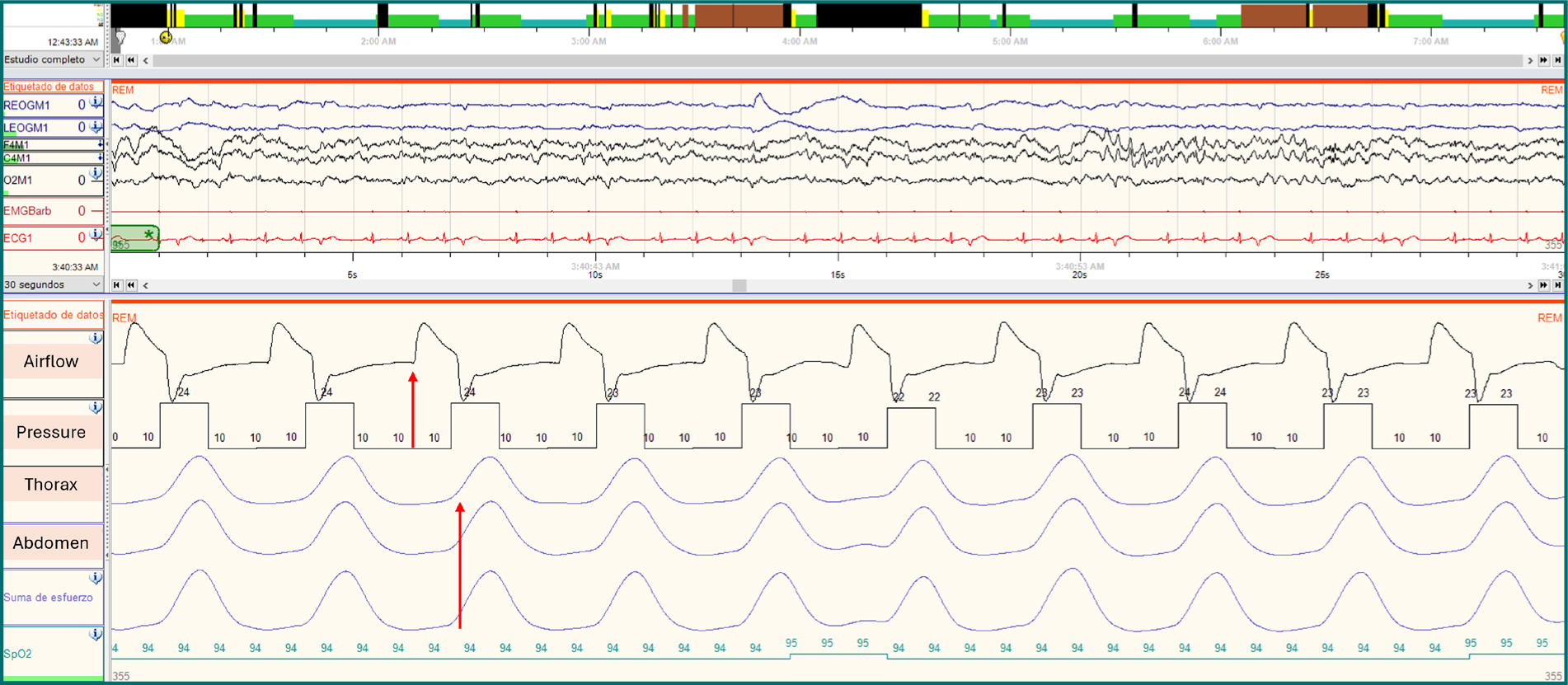
Figure 4. Polysomnography under non-invasive ventilator showing temporal delays between ventilator airflow and pressure signals (red arrow) and thoraco-abdominal effort at a 30 s screen (red arrow).
General evidence
PSG enables a direct assessment of the interactions between the patient and the ventilator during overnight use of NIV, as well as an evaluation of sleep quality and fragmentation. Patient-ventilator asynchrony denotes a mismatch between the patient’s neural inspiratory effort and the ventilator’s mechanical inspiratory phase. Observational studies have reported frequent PVA in individuals using nocturnal NIV after daytime titration,18 with improvements observed following adjustments to ventilator settings.19 Higher levels of PVA have been linked to more frequent EEG arousals during sleep,20 reductions in REM sleep and overall sleep efficiency,19 impaired nocturnal gas exchange,18 and decreased tolerance of NIV.21 Consequently, polysomnographic titration of NIV has been advocated, although few controlled trials have evaluated this approach.
As has been explained in this review, although polysomnographic titration of NIV is the gold standard of care, there is scarce evidence on its physiological and clinical benefits for naive users. The first randomized clinical trial with that purpose was conducted in 60 naive patients diagnosed with CHRF due to motor neuron disease (MND), neuromuscular disorders, obesity-hypoventilation syndrome (OHS), restrictive thoracic disorders, and chronic obstructive pulmonary disease (COPD)/obstructive sleep apnea (OSA).22 PVA event rates (mainly ineffective efforts) were reduced in those who underwent PSG-assisted setting optimization (PSG: 26 events/h [12–68] vs. control: 41 events/h [28–182], p = 0.046). Mean difference in average daily NIV use in those initially non-adherents to NIV (< 4 h/day) was more than one and a half hours higher in the PSG group versus the control group (PSG 95 [29–161] min vs. control −23 [−86 to 39] min). In a subset analysis of those with MND, PSG titration led to a higher proportion of initially non-adherent patients becoming adherent compared with controls. We have also published a recent crossover trial in 26 naive patients with CHRF due to different underlying diseases (n = 13 COPD, n = 8 neuromuscular disease [NMD], n = 3 OHS, and n = 2 chest wall disorders) comparing in-lab PSG manual titration accordingly AASM titration guidelines to an automatic adjustment by “learn target” procedure in intelligent volume-assured pressure support with automatic EPAP (IVAPS AE) mode.23 As Hannan et al. reported, despite the expertise manual titration of fixed pressures of ventilation, we did not found differences in sleep architecture, stages, sleep disruption, nocturnal or diurnal gas exchange during the attended laboratory PSG between manual titration and automatic mode. We did find a significant improvement for both modes in sleep disruption measured by arousals index, as is expected due to correcting hypoventilation. Patel et al.24 published a purpose of implementation of a single-night PSG split-titration using automatic volume-assured pressure support (AVAPS) in 27 patients with NMDs. They began titration in fixed pressure and spontaneous-timed (ST) mode, increasing IPAP and EPAP, also following AASM recommendations, and when mean SpO2 ≥ 90% was achieved, including REM sleep, ST mode was switched to AVAPS mode. The AVAPS titration continued, then applying the estimated average tidal volume value obtained during the ST mode titration. Although the design in a single-night PSG differs from our full night titration and as well the algorithm of VAPS from Philips and iVAPS from ResMed, our results were in agreement with theirs, showing higher average inspiratory pressures and tidal volumes in automatic VAPS mode compared to fixed pressure ST mode to achieve similar correction of nocturnal hypoventilation, and when patients were asked to the preference of NIV mode, they tended to prefer ST mode.
UA obstructive events related to NIV and/or type of mask
NIV is primarily applied during sleep, a period characterized by marked changes in respiratory mechanics and UA patency. Moreover, NIV itself may induce undesirable respiratory events, therefore a baseline sleep study before ventilation initiation would be desirable to identify preexisting OSA and to differentiate them from newly incurred.6 We reported a high number of hypopneas/hypoventilation in patients studied without suspicion of OSA.23 In patients with CHRF, this has been related to instability of the UA and/or decrease in ventilatory drive, leading to a reduction in airflow and belt signal amplitudes without phase opposition that could also be scored as hypopneas. Thus, PSG under ventilation enables precise characterization of UAO events and patient-ventilator interaction, particularly for events induced or modulated by NIV and/or mask interface choice. PSG uniquely discriminates obstructive events with or without ventilatory drive, detects sleep-specific phenomena, and guides targeted adjustments in EPAP, interface selection, and ventilator settings. Table 1 summarizes evidence supporting PSG titration for UAO interpretation and correction.
Table 1. Evidence supporting PSG titration for UAO events interpretation and correction
| Clinical problem | PSG-based phenotype | Association with mask type | Clinical advantage of PSG-guided titration |
|---|---|---|---|
| Treatment-induced UAO | New or worsened obstructive events appearing after NIV initiation, absent or less frequent on baseline sleep study | Predominantly reported with oronasal masks, especially in coexisting OSA and neuromuscular disease | Identifies NIV-induced obstruction and prevents misclassification as residual or untreated OSA |
| UAO without reduction in ventilatory drive | Preserved or increased inspiratory effort with flow reduction/limitation during pressurization | Occurs with both mask types; pressure-interface interaction critical | PSG distinguishes UAO from central events or asynchrony, guiding EPAP and interface adjustments |
| UAO with reduced ventilatory drive | Obstruction during ventilator-controlled breaths with absent or minimal respiratory effort | Less dependent on mask type | Enables correct event classification and avoids inappropriate escalation of pressure support |
| Mask-induced airway collapse (tongue or pharyngeal obstruction) | Obstructive events temporally linked to inspiratory pressure delivery | More frequent with oronasal masks, possibly due to posterior tongue displacement | Allows attribution of obstruction to interface rather than disease progression |
| Insufficient EPAP leading to residual obstruction | Persistent obstructive events despite adequate pressure support | Particularly relevant when using oronasal masks | PSG enables stepwise EPAP titration under sleep conditions to restore airway patency |
| Leak-related UAO and flow artefacts | Apparent obstructive events coinciding with major unintentional leaks | Nasal masks associated with higher nocturnal leak burden; oronasal masks reduce leak but may promote obstruction | PSG differentiates true UAO from leak-related artefacts overlooked by ventilator software |
| Accuracy of estimated AHI from BIS | Over/underestimation of AHI-BIS compared to AHI manually scored (leaks/UA events without increased UA resistance as in hypopneas/hypoventilation) | Occurs with both mask types. Related to device proprietary algorithm | PSG confirms true residual AHI regardless of the type of respiratory event |
PSG: polysomnography; UAO: upper airway obstruction; NIV: non-invasive ventilator; OSA: obstructive sleep apnea; EPAP: expiratory positive airway pressure; AHI: apnea-hypopnea index; BIS: built-in software; UA: upper airway.
Current evidence of PSG role relying on underlying diseases
ALS
Sleep disturbances affect up to 70% of ALS patients and were traditionally attributed to respiratory muscle weakness. However, evidence from early-stage ALS shows significant sleep architecture alterations despite preserved respiratory function, indicating central sleep-wake dysregulation. Hypothalamic dysfunction may underlie these disturbances and contribute to non-motor features such as autonomic dysfunction, hypermetabolism, and weight loss. In addition, impaired deep non-rapid eye movement sleep may disrupt glymphatic clearance, suggesting a potential role of sleep disturbances in ALS pathophysiology and progression. Although OSA prevalence varies considerably across studies, ranging from 17% to 76%25 is consistently higher than in the general population, with some estimates suggesting 50–80% of ALS patients exhibit OSA features.26,27 This heterogeneity reflects differences in disease stage at assessment, diagnostic criteria (apnea-hypopnea index [AHI] thresholds, type of sleep study), patient phenotype (bulbar versus spinal onset), and methodological approaches. The increased OSA burden is attributed to disease-specific factors, including progressive oropharyngeal muscle weakness from bulbar involvement and heightened REM sleep vulnerability when physiological muscle atonia further compromises respiratory control. Although AHI represents the cornerstone metric for OSA diagnosis and severity classification, one of the most striking limitations of AHI in ALS is its counterintuitive decline as the disease progresses attributable to: (1) progressive respiratory muscle weakness: as patients lose strength, they become less capable of generating the negative intrathoracic pressures required for UA collapse and the subsequent breathing efforts that define apneas and hypopneas; and (2) progressive weight loss, that contribute to the consequently replacement of the obstructive component by hypoventilation.28
Thus, PSG-guided titration of NIV provides a comprehensive and individualized method to optimize ventilatory support in patients with ALS. Evidence from multiple observational studies and randomized controlled trials supports its clinical relevance across several domains, including sleep quality, nocturnal gas exchange, patient-ventilator synchrony, respiratory events, adherence, and long-term outcomes.
PSG titration facilitates detection and correction of nocturnal hypoventilation, which often precedes daytime ventilatory failure in ALS. Early studies demonstrated significant improvements in sleep architecture and reductions in nocturnal hypercapnia and desaturation following meticulous PSG-guided titration, especially in nonbulbar ALS patients.29 These improvements were associated with enhanced subjective sleep quality and quality of life. To note, the methodology of PSG titration in that study was based on a prospective procedure in which patients were admitted to the sleep lab for four consecutive nights and not on a single in-lab PSG standard titration. Patients underwent to a first diagnostic-PSG night, the day after, IPAP was titrated in S mode during a nap to reach a tidal volume of 6 mL/min/kg ideal body weight, and in the morning of the 3 consecutive days, PSG were analysed and NIV settings were adjusted according to nocturnal PtcCO2, oxygen saturation and occurrence of respiratory events, napping each afternoon for 1 h to get accustomed to the new settings. After 1 month of NIV, patients were readmitted for monitoring NIV-PSG under the settings previously applied. The rationale for that procedure probably is related to the above mentioned difficulty and level of expertise required to perform a PSG titration, that lead to the clinicians to interpret during the day the findings on the monitoring PSG under NIV, introducing thereafter the new settings and titrating during a nap-PSG, instead of during a full night PSG titration.
Persistent PVA is common in ALS and may remain unrecognized without PSG. Vandoorne et al.30 in a second study found high rates of PVA even after titration. They scored PVA (ineffective efforts, double triggering, auto triggering, and premature cycling), leaks, and respiratory events. The most prominent PVA were ineffective efforts, with a significant higher ratio in non-bulbar patients compared with bulbar ones at discharge (43 [15–90] vs. 9 [4–31]/h sleep, p < 0.05) but disappearing after 1 month (21 [11–43] vs. 19 [10–40]/h sleep). Regarding leaks, although the proportion of sleep affected by leaks did not differ significantly between bulbar and nonbulbar patients or between assessment time points, the presence of leaks was consistently associated with increased PVA. Observing each type of asynchrony separately, no differences were found comparing asynchronies with or without leak, except for ineffective efforts in the nonbulbar patients after 1 month (56 [22–101]/h sleep during leak vs. 18 [10–25]/h sleep without leak; p < 0.05). Finally, the amount of UA respiratory events was negligible in the present study. Thus, even after optimized polysomnographic titration, monitoring PSG demonstrated persistent PVA and unintentional leaks that, despite minimal impact on sleep architecture, may negatively influence long-term tolerance and effectiveness of NIV.
Several randomized trials comparing specific NIV modes during PSG titration emphasize its relevance for guiding mode selection. In a crossover trial, Vrijsen et al. using the same titration procedure previously described, found that ST mode provided superior nocturnal gas exchange and fewer ineffective efforts and respiratory events, both obstructive and central/hour sleep than spontaneous (S) mode, although some patients still benefited from S mode (better sleep efficiency and arousal/awakening index).31 Similarly, Crescimanno et al.32 demonstrated also in a crossover randomized study that pressure-controlled ventilation improved sleep structure and reduced asynchronies, being predominantly ineffective efforts and underassistance, compared with volume-controlled ventilation, despite comparable blood gas correction, highlighting the limitations of relying solely on daytime titration or gas exchange metrics. Patients with ALS may be more prone to develop glottic events under NIV than healthy individuals. Such events have been reported, especially in subjects with pseudobulbar dysfunction33 or in subjects with spinal onset disease with early involvement of the vagus nerve.34 Glottic events have been attributed to laryngeal dysfunction and altered upper-airway reflex regulation and are exaggerated by high pressure or flow.35 In subjects with ALS, when using pressure-controlled ventilation, Georges et al.36 reported a remarkably high prevalence of abnormal respiratory events (45%). However, Sancho et al.33 during volume ventilation, reported a low prevalence of these events (10%). These data were consistent with Crescimanno results, which showed, despite residual events were scarce for both modes, fewer abnormal respiratory events under VC mode. A switch from pressure to volume ventilation may be considered if a high number of these events is observed in the pressure mode.
PSG titration also contributes to long-term adherence and clinical stability. Studies show that NIV adherence is strongly influenced by comfort and synchrony, both of which can be optimized through PSG-based adjustments.29,32 Despite the benefits shown from laboratory-based sleep studies, they are not used by all groups who treat respiratory failure in ALS. There is an ongoing randomized clinical trial14 to test the efficacy of the implementation of a PSG-assisted NIV titration in the commencement of the therapy. Besides, it will be addressed a new proposal to titrate ventilation in ALS patients. The authors hypothesise that the PSG intervention will improve synchrony, leading to greater NIV usage and consequently, better survival as previously observed in ALS cohorts using optimized NIV.
Collectively, these findings demonstrate that PSG-guided NIV titration in ALS provides clinically meaningful advantages by enabling precise adjustment of ventilator settings, identification of asynchronies, optimization of sleep quality and gas exchange, and support for long-term adherence contributing to survival benefit.
COPD
The coexistence of COPD and OSA, commonly referred to as the overlap syndrome, has important diagnostic and therapeutic implications in patients with stable CHRF candidates for long-term NIV. Epidemiological studies have reported prevalence estimates ranging from 0.5% to 39% and up to 65%.37,38 Importantly, patients with overlap syndrome experience more severe nocturnal oxygen desaturation and sleep fragmentation compared with those with COPD or OSA alone, consistent with synergistic pathophysiological interactions.39
Direct evidence comparing an OSA screening strategy with no screening in hypercapnic COPD patients initiating NIV is lacking.40 Nevertheless, several observational studies suggest that OSA identification and continuous positive airway pressure (CPAP) treatment in COPD patients are associated with improved outcomes. Marin et al. demonstrated significantly higher mortality in untreated overlap syndrome compared with COPD alone.41 Similarly, improved survival and reduced hospitalizations with CPAP have been reported in hypercapnic overlap patients, along with improvements in exercise capacity and functional status.42 Given the absence of direct outcome data, the automatic transfer switch (ATS) panel highlighted that identifying OSA before NIV initiation may alter clinical management in several ways. Some patients may require CPAP alone rather than NIV if OSA is the dominant cause of hypercapnia, whereas others may benefit from optimized NIV titration, particularly with higher EPAP settings to prevent UA collapse. In conclusion, despite very low certainty evidence, the ATS guideline suggested screening for OSA before initiating long-term NIV in patients with chronic stable hypercapnic COPD.40
The optimal method for initiating and titrating long-term NIV in stable hypercapnic COPD patients remains uncertain. Owing to the high prevalence and clinical implications of the overlap syndrome described above, PSG can facilitate precise EPAP adjustment to prevent UAO while targeting the lowest effective pressure support during formal in-lab titration. However, evidence demonstrating clinically meaningful benefits from this approach is limited. Only two randomized controlled trials have compared PSG-based titration with alternative NIV initiation strategies. Hannan et al. found no significant differences in mortality, quality of life, PVA, or adverse effects between PSG-titration and daytime titration with sham PSG, although the study population was heterogeneous and only indirectly applicable to COPD.22 Similarly, Patout et al. reported no clear advantages of PSG-guided titration compared with a nurse-led protocol in a small pilot trial involving patients with COPD and OSA.43 Pooled analyses did not demonstrate improvements in NIV adherence or PaCO2 reduction.40 In addition, aggressive overnight correction of hypercapnia may be undesirable and has been associated with potential physiological risks, including glottic closure and metabolic disturbance.44 In summary, current evidence does not support routine in-lab PSG to determine long-term NIV settings in COPD. Alternative approaches, including outpatient or home-based initiation with longitudinal adjustment, appear feasible and appropriate for most patients, reserving PSG for selected cases with persistent hypoventilation, UAO events, or unsolved PVA after step-by-step evaluation.
Another potential role of PSG studies in this population is the expiratory flow limitation (EFL) assessment with novel automatic ventilation modes that continuously titrate EPAP to the lowest value to abolish tidal EFL (EFLT). Two recent studies in which nocturnal EFLT was continuously measured using the forced oscillation technique under NIV offer important nuance that may help refine indications for advanced or PSG-associated titration strategies. Zannin et al.,45 in a randomized cross-over study demonstrated that auto-titration of EPAP to abolish EFLT significantly reduced ineffective efforts and decreased time spent in nocturnal hypercapnia compared with fixed-EPAP, although without significant differences in mean TcCO2 or oxygenation. These findings reveal that night-to-night variability in EFL can influence optimal EPAP requirements and that dynamic EPAP adjustment may improve patient-ventilator synchrony during sleep.45 Similarly, McKenzie et al.46 in an observational study confirmed that EFL fluctuates markedly throughout sleep, varies independently of sleep stage and body position, and frequently diverges from values obtained during supine wakefulness. Automated EPAP titration effectively abolished EFLT for most of the night and was well tolerated over 2 weeks of home use, highlighting the limitations of relying solely on static daytime measurements. These data suggest that traditional fixed-EPAP NIV setups may not adequately address the dynamic nocturnal mechanics of COPD. In summary, PSG may still have a selective role in NIV titration for COPD, particularly in patients with: (1) suspected or confirmed OSA, where EPAP requirements are driven by UAO; (2) marked intranocturnal variability in mechanics, such as prominent or unstable EFL; (3) persistent hypercapnia or PVA despite standard outpatient titration. Nonetheless, outside these contexts, routine PSG-based titration remains unsupported by current guidelines.
Obesity hypoventilation syndrome
PSG has historically played a central role in the diagnosis and titration of positive airway pressure therapies in OHS, a condition characterised by chronic daytime hypercapnia in obese individuals, frequently associated with severe OSA. Early evidence supporting PSG titration in OHS comes from studies such as the Respirology trial by Arellano-Maric et al.,47 which demonstrated that PSG-guided autotitrating CPAP could be safely used to reassess stable OHS patients previously treated NIV. In this multicentre interventional study, overnight PSG combined with transcutaneous capnography was used to evaluate the physiological response to auto-CPAP before conversion to fixed CPAP at home. Approximately 70-80% of patients maintained normocapnia after switching, without deterioration in sleep architecture, gas exchange, lung function, or health-related quality of life. Importantly, PSG served a gatekeeping role, allowing identification of CPAP responders and exclusion of patients with persistent nocturnal hypoventilation, highlighting its value in safely optimising treatment modality selection in selected, stable OHS populations.
As it was mentioned above, more recently, randomised controlled trials addressing the necessity of in-lab PSG titration for NIV initiation were focused on OHS populations. The pickwick titration trial by Sánchez-Quiroga et al.48 compared fully auto-adjusted NIV (volume-targeted pressure support with auto-EPAP) to conventional manually titrated NIV guided by in-lab PSG. Over 12 months of follow-up, auto-adjusted NIV proved non-inferior to PSG-titrated NIV in reducing daytime PaCO2, improving sleep-disordered breathing, symptoms, quality of life, and healthcare utilisation. This trial provides robust evidence that, in stable ambulatory OHS patients, long-term clinical outcomes do not depend on PSG-based NIV titration, supporting a paradigm shift towards simplified initiation pathways and selective rather than routine PSG use. Similar conclusions emerge from the OPIP trial by Murphy et al.,49 although PSG was used in the inpatient arm to guide titration, outpatient autotitration achieved comparable improvements in gas exchange, sleep-disordered breathing, and quality of life, with no clinically relevant differences in short-term outcomes. Notably, outpatient pathways were not inferior from a safety perspective; however, outpatient AVAPS AE mode was no longer cost-effective due to the higher number of interventions required, both for ventilator settings adjustments and for non-scheduled visits. Taken together, these recent studies indicate that PSG titration in OHS has evolved from a universal requirement to a targeted tool. PSG remains crucial in specific scenarios: defining OHS phenotype at baseline (with or without severe OSA), identifying CPAP failure, assessing persistent nocturnal hypoventilation, and guiding treatment transitions (e.g., NIV to CPAP). However, for stable ambulatory patients and given the rising prevalence of OHS, particularly when modern autotitrating NIV or CPAP devices are available, PSG titration does not appear essential for achieving optimal long-term outcomes.
Conclusion
PSG offers unparalleled insight into the complex interactions between sleep, respiration, and non-invasive ventilation, allowing accurate identification of UA obstructive events, patient-ventilator asynchronies, and interface-related complications that frequently remain undetected with daytime assessments or ventilator-embedded monitoring alone. The accumulated evidence reviewed here supports PSG-guided NIV titration as a valuable and clinically relevant tool in selected patient populations, notably those with ALS, in whom optimization of synchrony, sleep quality, and adherence is critical, and on coexisting OSA, previously diagnosed by screening PSG or due to high clinical suspicion. Conversely, in stable patients with COPD or OHS, current data do not justify routine laboratory-based PSG titration, as less resource-intensive strategies yield comparable physiological and clinical outcomes, particularly in light of recent advances in automatic algorithms for autotitrating EPAP and EFL. Performing a monitoring or titration nap PSG in place of a full-night PSG may represent a feasible and efficient strategy for incorporating this procedure into routine clinical practice. Future research should focus on refining patient selection criteria, improving integration of advanced monitoring technologies, and clarifying the long-term impact of PSG-guided strategies on outcomes beyond gas exchange, including quality of life, cost-effectiveness, and survival.
Acknowledgments
The author would like to thank the editors for the kind invitation to prepare this review.
Funding
None.
Conflicts of interest
None.
Ethical considerations
Protection of human subjects and animals. The authors declare that no experiments on humans or animals were performed for this research.
Confidentiality, informed consent, and ethical approval. This study does not involve personal patient data, medical records, or biological samples, and does not require ethical approval. SAGER guidelines do not apply.
Declaration on the use of artificial intelligence. The authors declare that AI was used for the creation of the figures.
