Points for clinical practice
– Obesity-related respiratory disturbances, including obstructive sleep apnea (OSA), are associated with an increased incidence of acute and chronic respiratory failure.
– Management of respiratory failure in these patients depends on the patient’s underlying situation and on sleep study results.
– In the acute setting, non-invasive ventilation (NIV) must be prioritized in all but the most severe cases.
– In steady-state situations, administrating non-invasive positive airway pressure by using continuous positive airway pressure (CPAP) or NIV is presently the treatment of choice. Both CPAP and NIV seem to be similarly effective, but they differ in their mechanisms, applications, and outcomes.
Introduction
Even if obesity is changing, its origins can be traced back to the origin of the species: survival of the fittest dictated eating for survival, storing energy, and being as inactive as possible to conserve energy. Maybe because of that, obesity history is a history of failure. We already know enough about the causes and how to manage it, and we don’t need a new scientific invention. Nevertheless, obesity prevalence is strikingly increasing and is nowadays a public health problem. In 2022, 890 million people were obese, that is, 16% of the world adult population. In the USA, the prevalence of obesity in adults aged > 20 increased from 22% in 1988% to 42.5% in 2018.1
Obesity, well-known as a cardiovascular risk factor, is also a “respiratory” risk factor. Obesity, especially “apple shape” type, can have profound adverse effects on the respiratory system. It can cause alterations in pulmonary function tests, respiratory mechanics, respiratory muscle strength and endurance, gas exchange, control of breathing, and exercise capacity. In this field obesity is a major contributor to respiratory morbidity and mortality.2–5
However, there are only two decades that obesity-related respiratory disorders began to take a place in medical publications. Nevertheless, fiction literature has largely anticipated (once again…) science: already in 1836, Charles Dickens drew in his novel.6 A profile that would be, certainly, the best characterization of an obese subject with respiratory disorders:
“… and on the box sat a fat and re-faced boy, in a state of somnolency…. the fat boy rose, opened his eyes, swallowed a huge piece of pie he had been in the act of masticating when he fell asleep… Joe-dams the boy he’s gone to sleep again.”
The post-humous paper of the Pickwick Club, Charles Dickens 1836
It was necessary to wait more than 150 years so that Bickelmann et al.,7 found a pathophysiological explanation to the “phenotype” of Joe, “this fat red-faced boy, that snores as his wait at table, becomes easily asleep and then stop to breath”, when they described apneas and alveolar hypoventilation in these subjects.
Obesity hypoventilation syndrome (OHS) is defined as the association of a body mass index (BMI) ≥ 30 kg/m2, daytime hypercapnia (PaCO2 ≥ 45 mmHg), and sleep-disordered breathing, after ruling out any other respiratory disorder which may cause alveolar hypoventilation.8 Prevalence of OHS is estimated at approximately 0.4% of the general population.8 It is frequently associated with obstructive sleep apnea (OSA). Prevalence of daytime hypercapnia in subjects with OSA syndrome and obesity increases with BMI and approaches 24% when BMI is > 40 kg/m.2,9,10
Gas exchange in obese individuals
ABGs are frequently altered in obese subjects, and abnormalities are directly proportional to BMI. Two main pathophysiological mechanisms may account for gas exchange abnormalities in these patients: V/Q inequality, responsible for isolated hypoxemia, and alveolar hypoventilation, responsible for the also called “obesity hypoventilation syndrome.”
Mechanisms of hypoxemia
Isolated hypoxemia has been reported in up to 30% of patients with severe obesity.11 Although usually mild, hypoxemia tends to be more pronounced in individuals with reduced lung volumes and is often observed predominantly in the supine position, with further worsening during sleep.12,13 The principal mechanism underlying this abnormality is an increased alveolar-arterial oxygen gradient (A-aPO2) resulting from ventilation-perfusion mismatch, particularly in the dependent lung regions. This phenomenon is attributable to two complementary mechanisms: increased perfusion of the pulmonary bases due to the hypervolemic state and elevated pulmonary blood volume associated with obesity, together with reduced ventilation caused by distal airway closure and alveolar collapse. Moreover, in patients with obesity-related hypoventilation, a proportion of the reduction in arterial oxygen tension is directly related to the concomitant increase in arterial carbon dioxide tension, reflecting alveolar hypoventilation. Hypoxemia is more prevalent and severe in individuals with massive obesity (BMI > 40 kg/m2), particularly in those with a central (“android” or “apple-shaped”) fat distribution, and appears to correlate with the degree of expiratory reserve volume (ERV) reduction. A decrease in ERV, while inspiratory capacity remains relatively preserved, leads to a reduction in functional residual capacity (FRC), which may fall below closing volume, thereby promoting distal airway closure and atelectasis. Hypoxemia is typically exacerbated in the supine position, where ventilation-perfusion mismatch is further increased.14
Mechanisms of alveolar hypoventilation in obesity: can’t breathe or won’t breathe?
While the prevalence of OHS is estimated at approximately 0.4% of the general population,8 alveolar hypoventilation is observed in approximately 8–20% of obese individuals referred for evaluation of sleep-disordered breathing (SDB), with higher prevalence among those with severe obesity.15,16 Compared with eucapnic obese subjects, hypercapnic patients exhibit more pronounced respiratory abnormalities, including reduced chest wall and respiratory system compliance, increased respiratory resistance, and greater impairment in pulmonary function tests, particularly lower ERV, total lung capacity, and vital capacity.17,18 They also demonstrate an altered breathing pattern characterized by increased respiratory rate and reduced tidal volume, while the inspiratory duty cycle (Ti/Ttot) remains largely unchanged. In addition, respiratory muscle strength and endurance are diminished, and ventilatory responses to chemical stimuli are markedly reduced or inappropriate.17,19 Consequently, the work and energy cost of breathing are substantially increased.18 In some studies, the work of breathing (WOB) was reported to be nearly 3 times higher than normal, while the oxygen cost of breathing approached tenfold normal values, potentially predisposing these patients to respiratory muscle fatigue.18 Early investigators considered obesity itself to be the primary cause of hypoventilation, based on observations that hypercapnic patients exhibited more severe restrictive ventilatory defects than those with uncomplicated obesity. However, the lack of a significant correlation between BMI and arterial carbon dioxide tension (PaCO2) suggests that additional pathophysiological mechanisms are involved.17,19 The development of hypoventilation in obesity is likely multifactorial and results from the interaction of several mechanisms (Table 1).17 Despite extensive investigation, it remains unclear why only a subset of morbidly obese individuals develops chronic alveolar hypoventilation.19 Two principal hypotheses have been proposed. The first, commonly referred to as the mechanical hypothesis, attributes hypoventilation to the increased respiratory load imposed by obesity.17,18 Reduced chest wall compliance and altered respiratory mechanics substantially increase the effort required to maintain adequate ventilation.17,18 This excessive mechanical burden may overwhelm inspiratory muscles, ultimately leading to hypoventilation. Although appealing, the “can’t breathe” hypothesis has several limitations. First, the severity of obesity correlates poorly with the degree of hypercapnia.19 Second, while obesity increases the elastic load of the respiratory system, thoracic compliance does not consistently correlate with BMI.19 Therefore, mechanical disadvantage alone is unlikely to fully explain the development of chronic hypercapnia.19 The second theory, known as the blunted ventilatory drive hypothesis, proposes that impaired central respiratory drive plays a central role in the pathogenesis of hypoventilation.17,20 According to this concept, respiratory centers fail to augment ventilatory output to the level achieved by non-hypercapnic obese individuals.18 Support for this hypothesis comes from studies demonstrating that although hypercapnic obese subjects often exhibit an increased basal ventilatory drive, their ventilatory response to hypercapnic stimulation – as assessed by P0.1 measurements during CO2 challenge – is diminished or inadequate.17 In practical terms, obese individuals require greater neural respiratory output to generate a given level of ventilation, and those unable to achieve this compensation may develop chronic hypercapnia.18 Using the CO2 rebreathing technique, Gilbert et al. demonstrated that hypercapnic obese patients differed from eucapnic obese subjects primarily by their reduced ventilatory responsiveness rather than by anthropometric or clinical characteristics.21 This observation strongly suggests an abnormality in automatic ventilatory control, particularly in CO2-related respiratory regulation.17 However, Chouri-Pontarolo et al., challenged the concept of a uniform ventilatory control defect among hypercapnic obese patients.22 They identified two distinct subgroups: one with blunted ventilatory responses and another with preserved responses. No significant differences were observed between these groups regarding age, BMI, polysomnographic parameters, or daytime arterial blood gases. Nevertheless, patients with impaired ventilatory responsiveness exhibited greater daytime sleepiness and more severe hypoventilation. Although abnormalities in ventilatory control are well recognized in obesity-related hypoventilation, it remains uncertain whether they are a cause or a consequence of the disorder.19 Chronic exposure to intermittent hypoxia and sleep fragmentation, particularly in patients with concomitant OSA, may attenuate central ventilatory responsiveness over time.18,15 Conversely, some investigators have reported that ventilatory responsiveness fails to normalize after substantial weight loss, suggesting that impaired respiratory drive may precede obesity.19 This observation has led to speculation regarding a possible genetic or familial predisposition.20
Table 1. Pathophysiological mechanisms potentially implicated in respiratory failure in the obese
| Hypoxemic respiratory failure |
| Increase of a-APO2 gradient secondary to V/Q mismatching (especially in the bases) Hypervolemic and hyperdynamic state (overperfusion) Airway closure and alveolar collapse |
| Hypercapnic respiratory failure Impaired or inappropriate ventilatory drive Decreased chest wall and lung compliance Inappropriate ventilatory load compensation Increased upper airway resistance and inspiratory threshold load Impaired response to elastic and resistive loads Increased WOB and oxygen cost of breathing Decreased ventilatory muscle strength and endurance Excito-contractile uncoupling (increasing of diaphragmatic EMG not translated to an increase of muscular strength) Respiratory muscle fatigue (peripheral and/or “central”?) |
| Diaphragmatic dysfunction secondary to: Increased adipose tissue deposition Mechanical disadvantage (inadequate length-tension relationship) Increased VCO2 Changes in respiratory patterns (more rapid and shallow breathing) Increased total respiratory resistance |
| Coexisting conditions Sleep apnea or upper airway resistance syndrome COPD |
| Aggravating conditions Supine position REM sleep |
An alternative interpretation has been proposed by some authors, who argue that hypercapnia may represent an adaptive physiological response rather than a direct consequence of impaired ventilatory control.18 According to this view, instead of sustaining the substantial energetic cost required to normalize PaCO2 in the presence of increased respiratory load, certain individuals tolerate elevated carbon dioxide levels to reduce the oxygen expenditure associated with excessive WOB.17 This concept has been summarized by the provocative suggestion that some obese patients “won’t breathe” rather than “can’t breathe.” In addition to obesity itself, two common comorbid conditions – chronic obstructive pulmonary disease (COPD) and OSA – may contribute to or exacerbate alveolar hypoventilation.15,20 Approximately 90% of patients with OHS have coexistent OSA, with nearly 70% having severe OSA (apnea-hypopnea index [AHI] ≥ 30 events/h).15,16 In the presence of OSA and reduced ventilation, CO2 accumulates during sleep, particularly when apneas are repetitive, reducing interapneic time and impairing CO2 clearance (see below). Similar to morbid obesity, COPD increases the WOB, alters respiratory mechanics, and reduces respiratory muscle efficiency.20 Consequently, coexisting COPD may significantly worsen hypercapnia in obese patients, and the degree of PaCO2 elevation has been shown to correlate inversely with forced expiratory volume in 1 s.20
OHS and OSA: a complex and controversial relationship
OSA is highly prevalent, and obesity is its major risk factor. Approximately two-thirds of patients with OSA are obese, whereas more than half of severely obese individuals (BMI > 40 kg/m2) present severe OSA.2 In obese subjects, particularly those with upper-body fat distribution, adipose tissue accumulates in the soft palate, tongue, and lateral and posterior pharyngeal walls. This excess soft tissue reduces pharyngeal lumen size, increases extraluminal pressure, alters upper-airway collapsibility, and, together with obesity-related reductions in lung volumes, promotes airway obstruction during sleep.24
OHS and OSA share numerous clinical features and frequently coexist. Both conditions are associated with excessive daytime sleepiness, fatigue, and morning headaches. Furthermore, 11–15% of obese patients with OSA exhibit daytime hypercapnia,9 while most hypercapnic obese patients have concomitant OSA. Hypercapnia is also more common in obese than in non-obese OSA subjects.23 Nevertheless, despite this overlap, the relationship between OHS and OSA remains incompletely understood. The absence of a standardized definition of OHS, particularly regarding its relationship with OSA, has generated substantial controversy.25,26
Some authors advocate, including OSA within the definition of OHS, emphasizing that only a minority of OHS patients lack significant OSA. Support for this concept comes from observations that demonstrated that continuous positive airway pressure (CPAP) therapy may normalize arterial blood gases in a subgroup of hypercapnic obese patients.27 However, neither these investigators nor others found a direct relationship between OSA severity, assessed by the AHI, and the occurrence of hypercapnia.27,28
The mechanisms through which OSA contributes to chronic hypercapnia remain uncertain. One hypothesis proposes that repetitive breathing against an obstructed airway impairs ventilatory load compensation, the physiological response required to maintain alveolar ventilation despite increased mechanical loads. Such impairment may result from respiratory muscle fatigue, inadequate recovery between apneic episodes, or diaphragmatic dysfunction secondary to recurrent post-apneic hyperventilation.29 Alternatively, hypercapnia may reflect a blunted ventilatory response to chemical stimuli, reducing compensatory ventilation during inter-apneic periods.
Ayappa et al., demonstrated that PaCO2 levels in hypercapnic OSA patients were directly related to the apnea/inter-apnea duration ratio.30 Maintenance of eucapnia during sleep requires equilibrium between CO2 accumulation during apnea and its elimination between events. Hypercapnia develops when post-apneic ventilation is insufficient to clear the accumulated CO2. Whether this blunted ventilatory response results from increased respiratory loading or represents an adaptive response to chronic hypoxia, hypercapnia, and sleep fragmentation remains unknown. Because arousals are more closely related to inspiratory effort than to oxygen desaturation, reduced inspiratory effort may decrease arousal frequency, favoring hypoventilation rather than recurrent apneas and hypopneas. Consequently, some authors have proposed that OHS represents an “end-stage” manifestation of OSA.29,31
Supporting this view, De Miguel et al., reported the emergence of obstructive apneas in patients initially diagnosed with OHS without OSA after correction of alveolar hypoventilation using non-invasive ventilation (NIV).32 They hypothesized that restoration of respiratory-center sensitivity unmasked pre-existing upper-airway obstruction. According to this model, patients may enter a vicious cycle in which apnea-induced hypoxemia and sleep fragmentation blunt ventilatory responsiveness, while abnormal respiratory mechanics impede restoration of post-apneic eucapnia, leading to worsening gas-exchange abnormalities.
In contrast, other authors reject the inclusion of OSA within the definition of OHS. They argue that OHS is defined as obesity-related alveolar hypoventilation occurring after exclusion of alternative causes of respiratory failure. Since OSA is itself a recognized cause of respiratory failure, it should be excluded when establishing the diagnosis. Clinical evidence also indicates that hypercapnia may develop in non-obese OSA patients. In the ANTADIR Observatory cohort, Laaban et al. reported PaCO2 > 45 mmHg in 7.2% of OSA patients with BMI < 30 kg/m2 after exclusion of COPD.9 Conversely, obesity alone, even in the absence of OSA, may lead to daytime hypercapnia.9
These findings suggest heterogeneity among hypercapnic obese patients. In some individuals, hypercapnia appears primarily related to SDB, as evidenced by its correction with CPAP therapy. In others, persistent hypercapnia despite effective CPAP, or its occurrence in the absence of OSA, underscores the independent contribution of obesity. Thus, the development of hypercapnia likely reflects the balance between nocturnal obstructive events and non-apneic hypoventilation.
Accordingly, obese hypercapnic patients may be divided into two phenotypes: those with coexisting severe OSA and those without severe OSA. To clarify this issue and address the ongoing terminological debate, alternative designations have been proposed, including obesity-linked hypoventilation (OLH),25 sleep hypoventilation syndrome (SHS),3 and OHS without OSA.33 These concepts encompass two clinical situations: (1) hypercapnia in obese patients without OSA or COPD (“pure” OLH, SHS, or OHS without OSA); and (2) persistent hypercapnia in OSA patients despite CPAP therapy (OLH or SHS associated with OSA, or OHS with OSA).3,25,33
Leptin and hypercapnia: the lost link?
The mechanisms explaining why alveolar hypoventilation develops in some obese individuals but not in others remain unclear. Leptin, an adipose-derived hormone, may provide part of the explanation. Acting through hypothalamic receptors, leptin is primarily involved in the regulation of body weight by suppressing appetite and increasing energy expenditure. More recently, evidence has suggested a role for leptin in the control of breathing, particularly in obesity.
Initial observations arose from animal models lacking the leptin gene. These animals exhibit marked abnormalities in respiratory control leading to chronic respiratory failure, particularly during sleep. They also show alterations in diaphragm myosin heavy-chain composition, reducing resistance to fatigue. Such changes may contribute to the reduced lung volumes and compliance observed in obesity and to the increased susceptibility to hypercapnia. Importantly, these abnormalities are reversed by leptin replacement therapy.34
These findings led to the hypothesis that leptin stimulates ventilatory drive in response to the increased respiratory load associated with obesity and that leptin deficiency could contribute to OHS. However, leptin deficiency is rare in human obesity; circulating leptin concentrations are generally elevated. Consequently, obesity has been proposed to represent a state of leptin resistance.35 Because leptin’s ventilatory effects are mediated through hypothalamic receptors, this resistance may result from impaired transport across the blood-brain barrier or defects in central leptin signaling, including receptor downregulation.
Several studies support an association between leptin abnormalities and altered ventilatory control in obesity.36–38 These observations highlight the importance of leptin-pathway dysfunction in the pathophysiology of obesity-related respiratory disorders and suggest that leptin may constitute a mechanistic link between OSA and hypercapnia.
Overall, it has been proposed that central nervous system leptin levels help maintain adequate alveolar ventilation and compensate for the increased ventilatory load of obesity. Alterations in leptin metabolism or signaling may therefore explain why some obese individuals develop hypoventilation whereas others do not, despite a similar respiratory burden. Hypercapnia is likely the consequence of complex interactions leading to a vicious cycle in which leptin may play a pivotal role (Fig. 1).
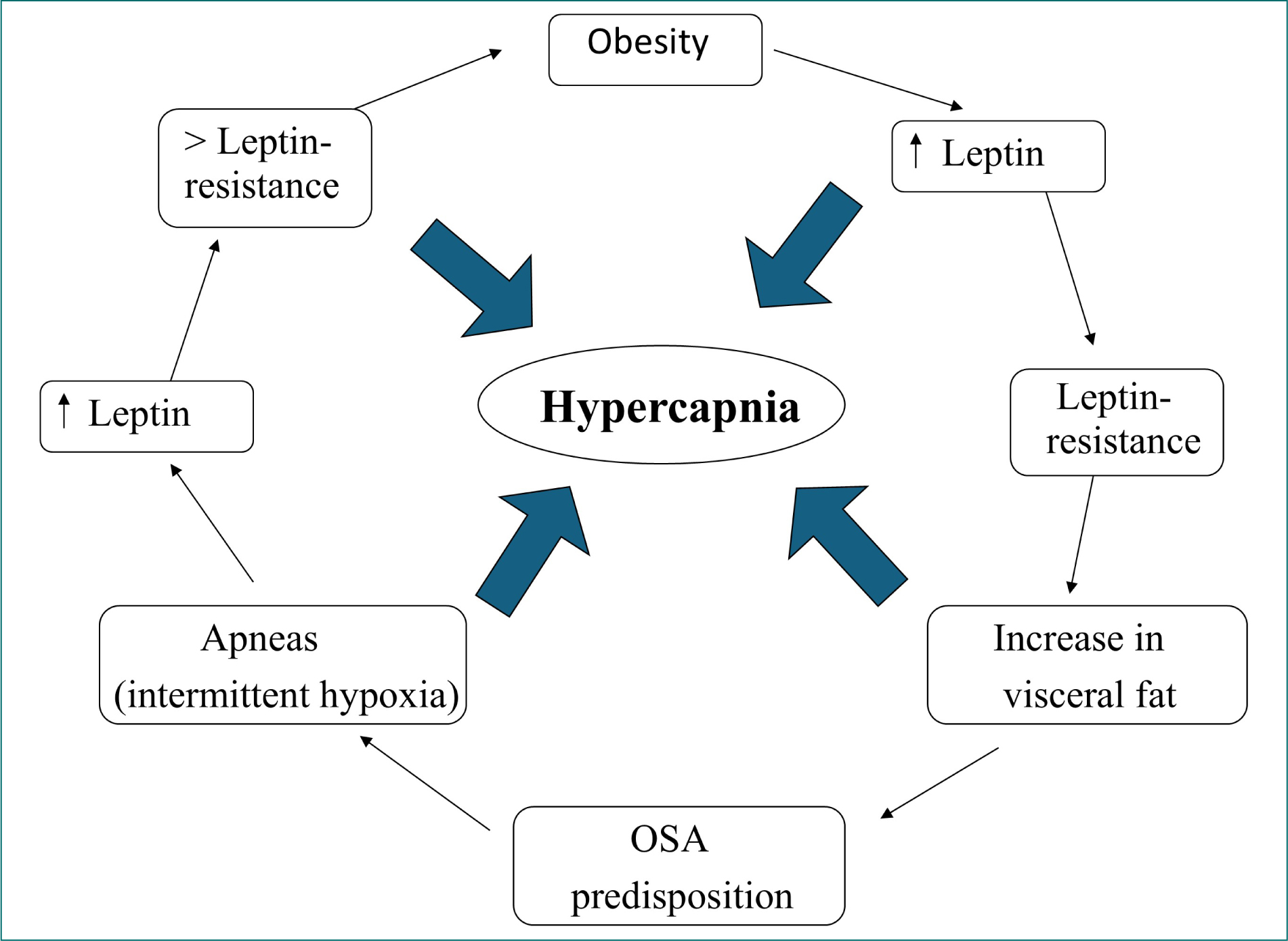
Figure 1. Leptin and hypoventilation in obese individuals (modified from Rabec et al.42).
Sleep and breathing in obesity
Respiratory failure in obesity is mainly driven by ventilation-perfusion (V/Q) mismatch and alveolar hypoventilation, both of which can worsen during sleep. The supine position further aggravates V/Q mismatch and promotes nocturnal hypoxemia. OSA and sleep-related hypoventilation, key mechanisms of obesity-related hypercapnia, are especially pronounced during sleep.16,39
Sleep studies reveal a wide spectrum of sleep-related breathing disorders in obese patients, including obstructive and central respiratory events, hypoventilation, and persistent gas-exchange abnormalities.33,39 OSA, defined by recurrent obstructive apneas and hypopneas, is highly prevalent in obesity and frequent in OHS. Central apneas may occur independently or after post-obstructive hyperventilation when PaCO2 falls below the apneic threshold.
Sleep hypoventilation is characterized by sustained nocturnal oxygen desaturation and, unlike isolated V/Q mismatch, is associated with nocturnal hypercapnia. Central hypoventilation reflects reduced ventilatory drive during sleep and is most evident in rapid eye movement (REM) sleep.22 During REM sleep, skeletal muscle atonia leaves the diaphragm as the main active respiratory muscle, and impaired diaphragmatic function combined with increased mechanical load can worsen hypoventilation.17
Obstructive hypoventilation corresponds to sustained hypoventilation due to partial upper-airway obstruction and represents an intermediate phenotype in SDB.33 Polysomnography, combining airflow and thoracoabdominal movement analysis, distinguishes central from obstructive mechanisms, which is essential for treatment decisions. Obstructive forms usually require CPAP optimization, whereas central hypoventilation often needs NIV.16
Excessive daytime sleepiness can occur in obese individuals even without documented SDB, suggesting roles for inflammation, metabolic dysfunction, and altered sleep architecture.39,40
OHS in clinical practice
OHS remains largely underdiagnosed. Patients typically present with severe obesity, daytime sleepiness, exertional dyspnea, and signs of right heart failure or cor pulmonale, but these are often misattributed to other conditions, such as COPD, heart failure, or depression, delaying diagnosis.16,41
Undiagnosed OHS is frequent in hospitalized obese patients. Studies show that 31% of patients with BMI > 35 kg/m2 have unrecognized hypoventilation, and nearly half of those with BMI > 50 kg/m2 present with daytime hypercapnia.3 However, BMI alone is not a reliable predictor of disease.
The pathophysiology of OHS is multifactorial, involving mechanical respiratory impairment due to obesity, altered ventilatory drive, SDB, and possible coexisting lung disease.41,42 OSA, COPD, and obesity-related hypoventilation often coexist, contributing to chronic respiratory failure and complicating diagnosis and management.
Diagnosis is commonly made either during acute-on-chronic hypercapnic respiratory failure – often triggered by infections or without clear cause – or during evaluation for SDB, when OHS may be missed if arterial blood gases are not assessed.16
OHS is associated with high morbidity and mortality, including pulmonary hypertension, secondary erythrocytosis, right ventricular dysfunction, and cor pulmonale.17 Pulmonary hypertension affects over half of patients in some series. Compared with eucapnic obese individuals, patients with OHS have higher rates of hospitalization, intensive care unit (ICU) admission, mechanical ventilation, and cardiovascular events, and untreated disease reduces long-term survival.3,16
Beyond clinical impact, OHS generates a significant healthcare burden through increased admissions and costs. Positive airway pressure (PAP) therapy improves gas exchange, quality of life, healthcare utilization, and survival.16,43 Systematic screening is therefore recommended in high-risk groups, such as patients with severe obesity, SDB symptoms, unexplained hypersomnolence, or chronic hypercapnia.16
Screening for respiratory abnormalities in the obese patient
OHS is frequently underdiagnosed and often identified only during acute respiratory failure (ARF). In patients on long-term NIV, nearly half are diagnosed during an acute hospital admission,44 highlighting delayed recognition.
Serum bicarbonate is a useful screening marker. According to ATS 2019 guidelines, a level < 27 mmol/L effectively rules out hypercapnia with a very high negative predictive value, while higher values should prompt further evaluation depending on pre-test probability.16 However, its positive predictive value is limited.
Because OHS risk increases in patients with OSA and BMI > 35 kg/m2, arterial blood gas analysis and sleep studies with transcutaneous CO2 monitoring should be performed in suspected cases or in those with mild daytime.9,10,16,43 SpO2 alone is not recommended for deciding when to perform ABG due to insufficient accuracy.
Screening tools, such as NoSAS or Berlin scores, can help estimate the probability of associated OSA and guide further diagnostic workup.
Therapeutical issues
Weight loss
Multimodal interventions combining caloric restriction, lifestyle modification, increased physical activity, and behavioral therapy constitute the first-line approach to obesity management; however, maintaining long-term effectiveness remains challenging.45 Pharmacological treatments and bariatric surgery have been shown to achieve substantial and sustained weight loss, with consequent significant improvements in obesity-related respiratory disorders.45,46 Benefits include substantial reductions in OSA severity, improvements in lung volumes and mechanics, enhanced gas exchange, and reduced airway resistance.47,48 The magnitude of benefit correlates with the degree of weight loss achieved. Instead, the ATS guideline recommends weight-loss interventions targeting sustained weight loss of 25–30% of actual body weight to achieve resolution of OHS.16 Weight loss of 7–17% through lifestyle modification produces 3–68% reduction in AHI, with greater baseline severity predicting larger absolute improvements.49 However, these improvements are reversible with weight regain, emphasizing the importance of sustained weight management in this population. Bariatric procedures, including Roux-en-Y gastric bypass and sleeve gastrectomy achieve 20–30% weight loss and produce substantial improvements in both OSA and pulmonary function.50,51 Meta-analyses show mean AHI reduction from 39.3 to 12.5 events/h following bariatric surgery, with greater reductions in patients with more severe baseline OSA.50 However, OSA persists in the majority of patients post-surgery despite significant improvement, necessitating continued monitoring and treatment.50 Introduced recently, GLP-1 receptor agonists (semaglutide, liraglutide, and dulaglutide) and dual GLP-1/GIP agonists (tirzepatide) achieve 15–20% weight loss at optimal dosing over 1 year.52 However, the long-term efficacy of these therapies remains unknown.
Ventilatory management in the obese patient
ARF
INVASIVE VENTILATION
The management of ARF in obese patients depends on disease severity. Patients with severe encephalopathy, severe pneumonia, or multiorgan failure should be admitted to the ICU, where prompt endotracheal intubation can be performed if required. In less severe cases, NIV, often combined with supplemental oxygen, remains the preferred first-line therapy.
Current indications for invasive mechanical ventilation (IMV) are mainly limited to contraindications or failure of NIV. Although these situations have become less frequent, repeated clinical reassessment is essential to avoid delayed intubation after prolonged ineffective NIV.
Obesity increases the risk of difficult intubation and extubation because of anatomical factors, such as a short neck, macroglossia, and excessive upper airway soft tissue. The Mallampati score remains a useful predictor of difficult airway management53 while a chin-anterior neck angle > 90° has also been associated with difficult intubation. Video laryngoscopy is recommended in critically ill obese patients to improve glottic visualization and first-pass success.54
Obesity-related reductions in FRC, especially in the supine position, markedly reduce oxygen reserves and shorten the safe apnea period before desaturation.55 Therefore, pre-oxygenation should be optimized using CPAP, NIV, or high-flow nasal oxygen (HFNO).56 Positive-pressure pre-oxygenation reduces severe hypoxemia by limiting alveolar collapse and increasing end-expiratory lung volume. In addition, obesity and OSA increase the risk of gastroesophageal reflux and pulmonary aspiration during airway management.57
Mechanical ventilation in obese patients is complicated by reduced respiratory system compliance, increased airway resistance, elevated pleural pressure, and a high propensity for atelectasis. Lung-protective ventilation strategies should therefore be systematically applied, including low tidal volumes, moderate-to-high positive end-expiratory pressure (PEEP), and recruitment maneuvers.58 Because airway plateau pressure may overestimate transpulmonary pressure, individualized PEEP titration is particularly important in obese patients.59
Ventilatory mode selection generally follows standard ICU practice, with volume-controlled ventilation preferred during deep sedation and pressure-support ventilation during weaning.
Obesity itself is an independent risk factor for post-extubation respiratory failure and reintubation, supporting the use of preventive respiratory support immediately after extubation.60,61 The high prevalence of OSA further increases extubation failure risk because of residual sedation, impaired upper-airway muscle activity, and delayed drug clearance.57
The role of early tracheostomy remains controversial. Some studies suggest that it may reduce the duration of mechanical ventilation, ICU length of stay, and ventilator-associated pneumonia in morbidly obese patients.61
Prophylactic non-invasive respiratory support has become a cornerstone of post-extubation management. NIV alone62,63 or alternating NIV and HFNO64 improves weaning success and reduces reintubation. A recent randomized trial further demonstrated a lower reintubation risk with NIV compared with HFNO in obese patients at intermediate risk of extubation failure.65 Accordingly, NIV should be initiated immediately after extubation in most obese patients, particularly those with OHS, chronic hypercapnia, or OSA. Many of these patients subsequently require long-term nocturnal ventilatory support, emphasizing the importance of early screening for SDB and OHS.
NON-INVASIVE VENTILATION
The efficacy of NIV as a first-line treatment for obese patients with severe hypercapnic acidosis was first demonstrated in the late 1990s. A pioneering study by Rabec et al., showed that NIV effectively prevented intubation in 39 of 40 obese patients with severe hypercapnic acidosis.66 While no randomized controlled trials directly compare NIV and IMV in obesity-related ARF, comparative studies indicate similar outcomes in ARF associated with obesity and acute exacerbations of COPD.67 Observational data also show acceptable outcomes for patients with obesity-related ARF treated with NIV.68
Care must be taken when extrapolating data from COPD to obesity-related ARF, as exacerbations of COPD have well-characterized presentations, while obese patients may present with ARF due to various pathologies, including respiratory infections and heart failure.67,69 Clinicians managing ARF in obese patients must identify and address the cause of decompensation. Management can occur in respiratory critical care units, ICUs, or general wards, depending on severity and the medical team’s NIV expertise.
When initiating NIV in obese patients, clinicians should consider factors indicating a higher failure rate, such as super obesity, pneumonia at presentation, and severe physiological abnormalities.70,71 Poor compliance with home PAP also predicts failure during acute admission.70 Although patients with risk factors for treatment failure can still receive NIV, it should occur in a setting capable of escalating to invasive ventilation if necessary. The outcomes for patients failing NIV without intubation can be poor, with failure rates as high as 50–100%.68,71
If escalation to IMV is needed, intubating obese patients poses technical challenges, with risks of life-threatening complications during intubation significantly higher in this population.72 In addition, obesity is associated with various non-respiratory comorbidities that can affect management and outcomes in critical care.73 However, when controlling for comorbidities, obesity itself does not appear to worsen outcomes for invasively ventilated patients and may even confer a survival benefit.74
Patients presenting with acute on chronic respiratory failure should be evaluated for long-term home PAP after resolving acute decompensation but before discharge.16 It remains unclear whether NIV or CPAP provides similar outcomes in this context. Expert consensus recommends discharging patients with NIV set to empiric settings, with a follow-up assessment within 3 months to consider long-term home NIV or transitioning to CPAP if appropriate.16 Some authors emphasize the importance of PSG recordings for follow-up after achieving stability.66
Management of chronic respiratory failure: CPAP or NIV?
PAP therapy is the cornerstone of treatment for SDB and chronic respiratory failure in patients with OHS. CPAP and NIV constitute the two principal therapeutic approaches. Although both modalities improve gas exchange and sleep-related breathing disturbances, they differ substantially in their physiological effects and clinical indications.
CPAP acts by providing continuous distending pressure throughout the respiratory cycle, thereby preventing upper-airway collapse and reducing obstructive respiratory events. Although CPAP does not directly augment ventilation, suppression of apneas and hypopneas may improve nocturnal and daytime gas exchange, leading to normalization of daytime PaCO2 in a subset of patients.31 Because more than 70% of patients with OHS have coexisting severe obstructive sleep apnea (OSA), CPAP alone may adequately correct respiratory failure in many individuals.75
In contrast, NIV provides active ventilatory assistance by delivering inspiratory pressure support in addition to expiratory PAP (EPAP). Pressure-targeted bilevel ventilators remain the most commonly used devices.75,76 Independent adjustment of inspiratory PAP (IPAP) and EPAP enables simultaneous treatment of upper-airway obstruction and alveolar hypoventilation. The pressure difference between IPAP and EPAP increases tidal volume, reduces respiratory muscle workload, and improves alveolar ventilation. NIV may be delivered in spontaneous (S), spontaneous-timed (ST), or timed (T) modes. Among these, the ST mode is most frequently used because it combines patient-triggered breathing with a backup respiratory rate. In stable OHS, Contal et al., demonstrated superior control of residual SDB with ST compared with spontaneous mode alone.77
The optimal initial treatment of patients with OHS associated with severe OSA remains debated. In contrast, NIV is generally considered the treatment of choice for patients with OHS without significant OSA.
Three randomized controlled trials compared CPAP and NIV in stable hypercapnic obese patients with severe OSA (mean AHI > 60 events/h).79–82 Follow-up ranged from 3 months to 2 years. Collectively, these studies demonstrated no significant differences between CPAP and NIV regarding adherence, improvement in daytime PaCO2 and PaO2, oxygen requirements, healthcare utilization, daytime sleepiness, or overall quality of life. Only one study reported superior subjective sleep quality and vigilance with NIV81 (Table 2).
Table 2. Summary of published randomized controlled trials comparing continuous positive airway pressure and noninvasive ventilation
| Reference | Population/study design | Settings/PS level | Outcomes | Main results |
|---|---|---|---|---|
| Piper, 200880 | 3 months, 36 patients BMI = 53 (8) kg/m2 PaC2= 50 (47-57) mm Hg |
EPAP 10 cm IPAP 16 cm S mode Mean PS: 6 cm |
Primary: PaCO2 improvement Secondary: QOL, compliance, vigilance, sleepiness |
ABG NIV > CPAP in more severe hypercapnic patients QOL NIV = CPAP Sleep quality, vigilance NIV > CPAP |
| Masa, 201981 | 2 months, then 2 years, 221 patients BMI = 45 (7,6) kg/m2 PaCO2= 50 (4,5) mm Hg |
EPAP 8,2 cm IPAP 19,7 cm ST mode Mean PS: 11.5 cm |
Primary Short term study: PaCO2 improvement Long term study: admission/year Secondary: QOL, compliance, 6MWT, symptoms |
ABG/QOL/admissions NIV = CPAP |
| Howard, 201782 | 3 months, 60 patients BMI = 54,9 (11,9) kg/m2 PaCO2 = 60,7 (13,5) mm Hg |
EPAP 11,9 cm IPAP 18,3 cm ST mode Mean PS: 6,4 cm |
Primary Treatment failure (admissions, persistent hypercapnia or non-adherence) Secondary QOL, sleepiness |
Admissions compliance/QOL NIV = CPAP ABG NIV > CPAP in more severe hypercapnic patients |
IPAP: inspiratory positive airway pressure; QOL: quality of life; BMI: body mass index; ABG: arterial blood gases; PS: pressure support; EPAP: expiratory positive airway pressure.
Modified from Rabec et al.17
Moreover, the long-term Pickwick study found no difference between CPAP and NIV in cardiovascular outcomes, hospitalization rates, mortality, or exercise capacity after 2 years of follow-up.81 These findings have been confirmed by subsequent guideline reviews and support the concept that CPAP and NIV provide comparable clinical benefits in many patients with OHS-OSA overlap.
Nevertheless, several factors limit the generalizability of these trials to routine clinical practice. First, all studies enrolled clinically stable patients, whereas NIV is frequently initiated following acute hypercapnic respiratory failure. In the French ANTADIR-GAVO2 registry, two-thirds of patients with OHS commenced home NIV after hospitalization for ARF.77 Furthermore, the prolonged recruitment period of the Pickwick trial suggests inclusion of a highly selected patient population.81
Second, baseline hypercapnia was relatively modest in these studies, with mean PaCO2 values around 50 mmHg. Only a minority of patients exhibited severe daytime hipercapnia.81 Some observational studies have identified milder hypercapnia and more severe OSA as predictors of successful treatment with CPAP alone.83,84 Consistent with this observation, Howard et al., reported that patients with baseline PaCO2 > 60 mmHg had an eight-fold higher risk of persistent hypercapnia during CPAP therapy than those with PaCO2 < 50 mmHg.79
Third, inspiratory pressure support levels used in the NIV arms were lower than those commonly applied in clinical practice. Mean pressure support ranged from 6 to 11.5 cmH2O,70,79,81 whereas studies specifically targeting correction of hypoventilation often employed higher pressures.82 Given the markedly reduced respiratory system compliance observed in severe obesity,85 insufficient pressure support may have limited the physiological effectiveness of NIV in these trials. Indeed, almost half of NIV-treated patients in the Pickwick study remained hypercapnic after 2 years.81
Toward a personalized therapeutic strategy
Recent evidence supports a more individualized approach to PAP therapy. Several studies have shown that patients initially treated with NIV who achieve normalization of daytime PaCO2 can subsequently be transitioned safely to CPAP without deterioration in blood gases, sleep quality, or health-related quality of life.86,87 These findings support a sequential strategy whereby NIV is used initially to correct severe hypoventilation, followed by simplification to CPAP once stable normocapnia is achieved.
From a pathophysiological perspective, hypercapnia in OHS may result predominantly from severe OSA, obesity-related ventilatory impairment, or a combination of both mechanisms. Distinguishing between these phenotypes is often possible only retrospectively, based on the response to treatment.66
Consequently, several experts advocate tailoring therapy according to both daytime PaCO2 and OSA severity.17,88–90 Patients with confirmed OSA and mild-to-moderate hypercapnia (PaCO2 < 50 mmHg) are reasonable candidates for an initial CPAP trial. Treatment efficacy should be reassessed after approximately 3 months using arterial blood gases, symptom evaluation, sleep quality, and cardiopulmonary outcomes. Persistent hypercapnia despite adequate adherence suggests ongoing obesity-related hypoventilation and should prompt transition to NIV90 (Fig. 2).
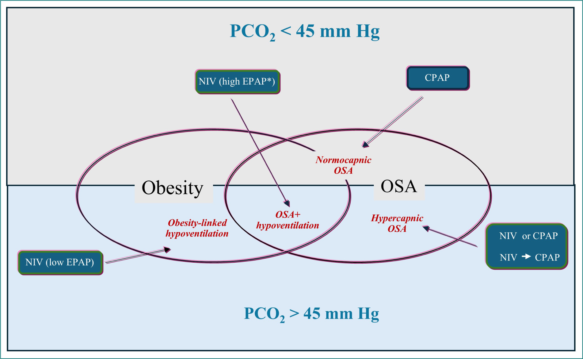
Figure 2. OHS-OSA relationships and pathophysiological-based therapeutic approach (modified from Rabec et al.17). OSA: obstructive sleep apnea; NIV: non-invasive ventilation; CPAP: continuous positive airway pressure.
Conversely, patients with marked daytime hypercapnia (PaCO2 > 50 mmHg), severe hypoventilation, or previous episodes of acute hypercapnic respiratory failure are more likely to benefit from NIV as first-line therapy. Once normocapnia is achieved, selected patients may subsequently be stepped down to CPAP86,87 (Fig. 3).
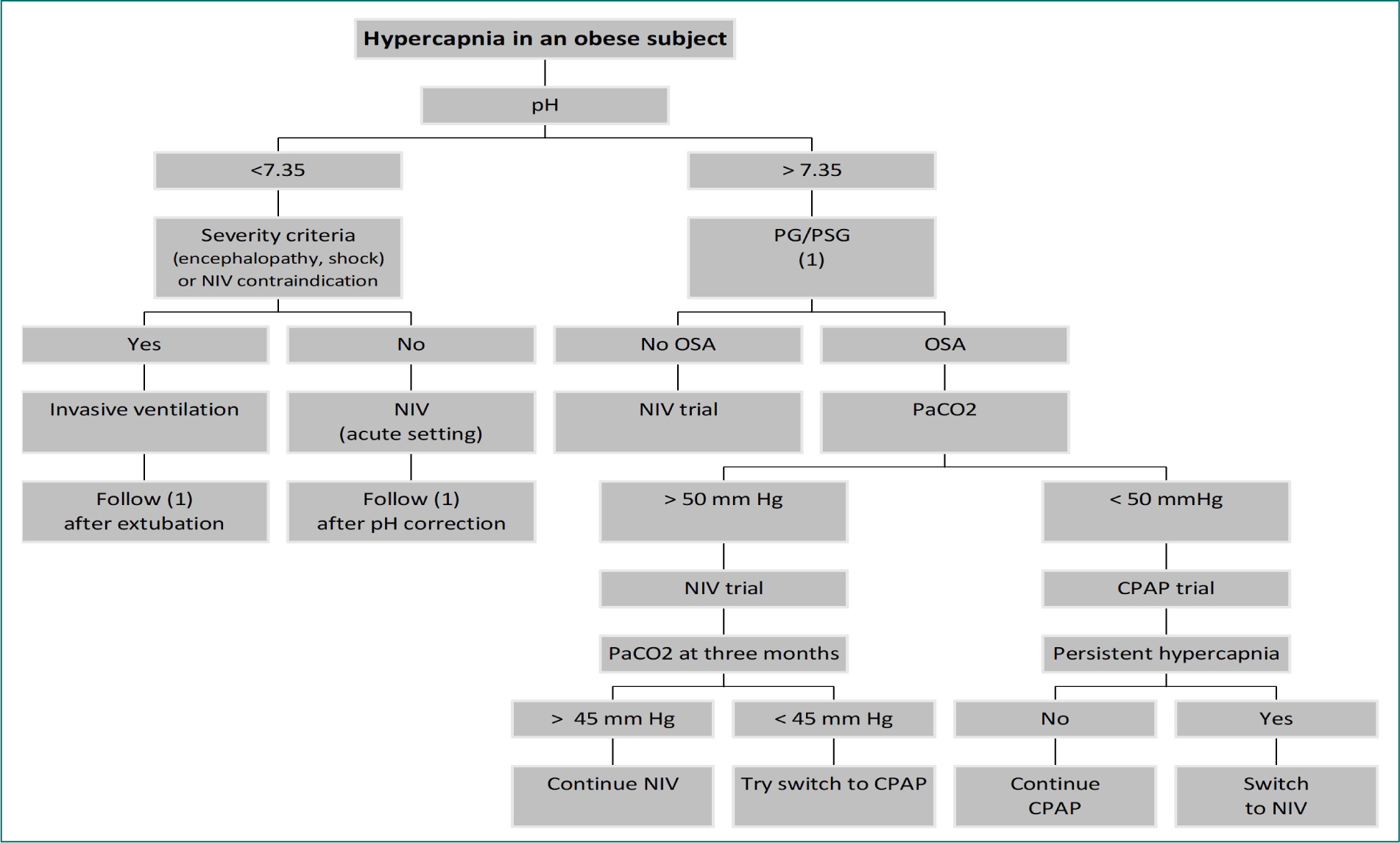
Figure 3. Proposed flow chart to ventilatory management of patients with obesity hypoventilation (modified from Rabec et al.17). OSA: obstructive sleep apnea, NIV: non-invasive ventilation; CPAP: continuous positive airway pressure; PG/PSG: polygraphy/polysomnography.
In a small subgroup of patients, adequate correction of hypoventilation cannot be achieved despite high levels of pressure support. These individuals may require alternative ventilatory strategies, including volume-targeted ventilation or, exceptionally, tracheostomy when non-invasive approaches fail or are poorly tolerated.
Role of hybrid modes of NIV
Pressure-limited bilevel ventilation remains the most widely used modality for long-term home NIV in patients with obesity-related respiratory failure.76,77 By delivering a pre-defined inspiratory pressure support, these devices provide effective leak compensation, improve patient comfort, and reduce respiratory muscle workload.91 However, fixed-pressure ventilation does not account for the substantial variability in respiratory system mechanics that occur during sleep, changes in body position, progression of underlying disease, or intercurrent respiratory events.92,93
These limitations are particularly relevant in obesity, where respiratory compliance and upper-airway mechanics may fluctuate considerably throughout the night. Supine positioning, sleep-stage transitions, and dynamic changes in upper-airway obstruction may significantly alter ventilatory requirements.94,95 Advances in ventilator technology have therefore led to the development of hybrid modes that combine the advantages of pressure-targeted ventilation with automated volume targeting and upper-airway management.
Volume-targeted ventilation
Volume-targeted modes were designed to maintain pre-defined target ventilation while preserving the comfort and leak compensation associated with pressure-limited NIV. These systems continuously adjust inspiratory pressure support to achieve a target tidal volume or alveolar ventilation. However, their performance depends on proprietary algorithms and estimated physiological parameters, which may vary between manufacturers and devices.96 Consequently, evidence obtained with one platform should not be extrapolated indiscriminately to others.
Several studies have demonstrated that volume-targeted NIV is safe and effective in chronic respiratory failure due to obesity, neuromuscular disorders, or COPD.82,97–99 Early investigations suggested improved nocturnal ventilation control in OHS compared with fixed pressure bilevel ventilation, although this benefit was frequently achieved through the delivery of higher mean inspiratory pressures rather than through intrinsic superiority of the ventilation mode itself.
For example, Storre et al., reported a greater reduction in nocturnal transcutaneous carbon dioxide levels with volume-targeted ventilation than with conventional bilevel NIV.99 However, this improvement was accompanied by significantly higher inspiratory pressures. Such pressure fluctuations may increase mask leaks, sleep fragmentation, and patient discomfort, potentially offsetting physiological benefits.
Subsequent studies using standardized titration protocols have challenged the notion that volume-targeted ventilation is intrinsically superior. Murphy et al. demonstrated that when pressure support levels were appropriately optimized in both treatment arms, volume-targeted and fixed-pressure NIV achieved similar control of nocturnal hypoventilation, daytime gas exchange, and treatment adherence.100 These findings suggest that careful titration may be more important than the choice of ventilatory mode itself.
Although some studies in COPD populations have reported modest improvements in device adherence with volume-targeted modes,96–99 available evidence has not demonstrated consistent superiority regarding daytime hypercapnia, quality of life, hospitalizations, or survival. Moreover, the comparable long-term outcomes observed with CPAP and NIV in OHS suggest that any potential advantage of volume-targeted over fixed bilevel NIV is likely to be modest.8,75
Auto-EPAP and closed-loop ventilation strategies
More recently, hybrid NIV systems incorporating automatic adjustment of expiratory PAP (auto-EPAP) have been introduced. These devices aim to simultaneously control upper-airway obstruction and alveolar hypoventilation by dynamically adapting ventilator settings throughout the night.44
Different technological approaches are used to estimate upper-airway patency. Some systems rely on airflow analysis, whereas others employ forced oscillation techniques (FOT).101 Flow-based methods allow breath-by-breath adaptation and have been extensively validated in CPAP therapy. However, their accuracy decreases in the presence of large leaks or high airflow rates, conditions frequently encountered during NIV.102 FOT-based systems may be less sensitive to leak-related artefacts but assess airway patency intermittently and may therefore fail to detect transient obstructive events or occasionally overcorrect pressure settings.103
Clinical studies conducted in patients with obesity-related respiratory failure have demonstrated that auto-EPAP strategies effectively control SDB while maintaining sleep quality and treatment tolerance.44 Polysomnographic evaluations showed no significant differences in sleep efficiency, arousal index, or wake time after sleep onset when compared with conventional bilevel NIV. Improvements in daytime PaCO2 and treatment adherence were also comparable between modalities.44
One potential advantage of auto-adjusting systems is the simplification of NIV initiation and titration. In a multicenter randomized study, Murphy et al., compared conventional inpatient NIV titration with outpatient initiation using an auto-EPAP volume-targeted device in patients with obesity-related respiratory failure.104 Both strategies achieved similar improvements in daytime hypercapnia, health-related quality of life, and healthcare costs, while reducing the time required to establish effective therapy. These findings suggest that selected patients may be safely initiated on NIV outside specialized inpatient settings.
Despite their increasing sophistication, available studies have not demonstrated clear superiority of hybrid modes over well-titrated conventional bilevel ventilation regarding clinically meaningful outcomes. Their effectiveness remains dependent on appropriate titration, careful interpretation of device data, and regular assessment of treatment efficacy. Hence, hybrid ventilation modes should not be considered self-adjusting solutions that eliminate the need for expert supervision.
Particular attention should be paid to target settings. Excessively wide pressure ranges, inappropriate target volumes, or inadequate pressure-support limits may result in persistent SDB and residual hypoventilation despite apparently satisfactory device performance.105 Furthermore, the accuracy of estimated ventilatory parameters may deteriorate substantially in the presence of large unintentional leaks.96
Conclusion
Obesity significantly affects respiratory mechanics and function, leading to obesity-related respiratory disturbances, such as OSA, which are linked to a heightened risk of both acute and chronic respiratory failure. The management of respiratory failure in these patients is contingent upon their underlying conditions and the results of sleep studies. In acute scenarios, NIV should be prioritized, as it represents the gold standard treatment in all but the most critical cases. For patients with stable chronic respiratory failure, non-invasive PAP is currently the preferred intervention for addressing SDB in stable OHS patients. While both CPAP and NIV demonstrate comparable efficacy, they differ in their mechanisms, applications, and outcomes. A tailored approach to OHS management is essential, recommending either NIV or CPAP based on the severity of hypercapnia and the presence and severity of OSA. Further research is necessary to delineate the optimal role of each modality in the management of patients with OHS.
Funding
None.
Conflicts of interest
None.
Ethical considerations
Protection of human subjects and animals. The authors declare that no experiments on humans or animals were performed for this research.
Confidentiality, informed consent, and ethical approval. This study does not involve personal patient data, medical records, or biological samples, and does not require ethical approval. SAGER guidelines do not apply.
Declaration on the use of artificial intelligence. The authors declare that no generative artificial intelligence was used in the writing or creation of the content of this manuscript.
